 Movie
Movie Controller


+ Open data

- Basic information
| Entry | Database: PDB / ID: 1l0i | ||||||
|---|---|---|---|---|---|---|---|
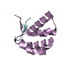
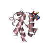
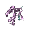
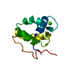
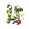
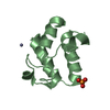
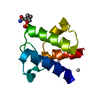
| Title | Crystal structure of butyryl-ACP I62M mutant | ||||||
 Components Components | Acyl carrier protein | ||||||
 Keywords Keywords | LIPID TRANSPORT / acyl carrier protein / acyl chain binding / fatty acid biosynthesis | ||||||
| Function / homology |  Function and homology information Function and homology informationlipid A biosynthetic process / lipid biosynthetic process / phosphopantetheine binding / acyl binding / acyl carrier activity / fatty acid biosynthetic process / response to xenobiotic stimulus / lipid binding / membrane / cytoplasm / cytosol Similarity search - Function | ||||||
| Biological species |  | ||||||
| Method |  X-RAY DIFFRACTION / SYNCHROTRON / MAD / Resolution: 1.2 Å X-RAY DIFFRACTION / SYNCHROTRON / MAD / Resolution: 1.2 Å | ||||||
 Authors Authors | Roujeinikova, A. / Baldock, C. / Simon, W.J. / Gilroy, J. / Baker, P.J. / Stuitje, A.R. / Rice, D.W. / Slabas, A.R. / Rafferty, J.B. | ||||||
 Citation Citation | Journal: Structure / Year: 2002 Title: X-ray Crystallographic Studies on Butyryl-ACP Reveal Flexibility of the Structure around a Putative Acyl Chain Binding Site Authors: Roujeinikova, A. / Baldock, C. / Simon, W.J. / Gilroy, J. / Baker, P.J. / Stuitje, A.R. / Rice, D.W. / Slabas, A.R. / Rafferty, J.B. #1: Journal: Acta Crystallogr.,Sect.D / Year: 2002Title: Crystallisation and preliminary X-ray crystallographic studies on acyl-(acyl carrier protein) from Escherichia coli Authors: Roujeinikova, A. / Baldock, C. / Simon, W.J. / Gilroy, J. / Baker, P.J. / Stuitje, A.R. / Rice, D.W. / Rafferty, J.B. / Slabas, A.R. | ||||||
| History |
| ||||||
| Remark 300 | Only the butryl, beta-mercaptoethylamine and phosphate moieties of the acylated 4' ...Only the butryl, beta-mercaptoethylamine and phosphate moieties of the acylated 4' phosphopantetheine group (PSR) attached to ACP were seen in the electron density. The rest of the 4' phosphopantetheine group was modelled in a stereochemically reasonable manner but omitted from the refinement process (occupancies set to 0.00). |
- Structure visualization
| Structure viewer | Molecule: MolmilJmol/JSmol |
|---|
- Downloads & links
-Download
| PDBx/mmCIF format | 1l0i.cif.gz | 55.3 KB | Display | PDBx/mmCIF format |
|---|---|---|---|---|
| PDB format | pdb1l0i.ent.gz | 38.9 KB | Display | PDB format |
| PDBx/mmJSON format | 1l0i.json.gz | Tree view | PDBx/mmJSON format | |
| Others |  Other downloads Other downloads |
-Validation report
| Arichive directory | https://data.pdbj.org/pub/pdb/validation_reports/l0/1l0iftp://data.pdbj.org/pub/pdb/validation_reports/l0/1l0i | HTTPS FTP |
|---|
-Related structure data


-Links
 PDBj
PDBj


- Assembly
| Deposited unit | 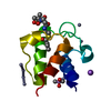
| ||||||||
|---|---|---|---|---|---|---|---|---|---|
| 1 |
| ||||||||
| Unit cell |
|
-Components
-Protein , 1 types, 1 molecules A
| #1: Protein | Mass: 8663.499 Da / Num. of mol.: 1 / Mutation: I62M Source method: isolated from a genetically manipulated source Source: (gene. exp.) |
|---|
-Non-polymers , 5 types, 172 molecules 


| #2: Chemical | ChemComp-NA /  Mass: 22.990 Da / Num. of mol.: 1 / Source method: obtained synthetically / Formula: Na Mass: 22.990 Da / Num. of mol.: 1 / Source method: obtained synthetically / Formula: Na | ||||||
|---|---|---|---|---|---|---|---|
| #3: Chemical | ChemComp-ZN /  Mass: 65.409 Da / Num. of mol.: 7 / Source method: obtained synthetically / Formula: Zn Mass: 65.409 Da / Num. of mol.: 7 / Source method: obtained synthetically / Formula: Zn#4: Chemical | ChemComp-CAC / |  Mass: 136.989 Da / Num. of mol.: 1 / Source method: obtained synthetically / Formula: C2H6AsO2 Mass: 136.989 Da / Num. of mol.: 1 / Source method: obtained synthetically / Formula: C2H6AsO2#5: Chemical | ChemComp-PSR / |  Mass: 428.438 Da / Num. of mol.: 1 / Source method: obtained synthetically / Formula: C15H29N2O8PS Mass: 428.438 Da / Num. of mol.: 1 / Source method: obtained synthetically / Formula: C15H29N2O8PS#6: Water | ChemComp-HOH / | Mass: 18.015 Da / Num. of mol.: 162 / Source method: isolated from a natural source / Formula: H2O |
-Details
| Has protein modification | Y |
|---|
-Experimental details
-Experiment
| Experiment | Method: X-RAY DIFFRACTION / Number of used crystals: 1 |
|---|
- Sample preparation
| Crystal | Density Matthews: 2.1 Å3/Da / Density % sol: 45 % | ||||||||||||||||||||||||||||||
|---|---|---|---|---|---|---|---|---|---|---|---|---|---|---|---|---|---|---|---|---|---|---|---|---|---|---|---|---|---|---|---|
| Crystal grow | Temperature: 290 K / Method: vapor diffusion, hanging drop / pH: 6 Details: PEG 4000, zinc acetate, sodium cacodylate, pH 6.0, VAPOR DIFFUSION, HANGING DROP, temperature 290K | ||||||||||||||||||||||||||||||
| Crystal grow | *PLUS Details: Roujeinikova, A., (2002) Acta Crystallogr., Sect.D, 58, 330. | ||||||||||||||||||||||||||||||
| Components of the solutions | *PLUS
|
-Data collection
| Diffraction | Mean temperature: 100 K |
|---|---|
| Diffraction source | Source: SYNCHROTRON / Site: SRS  / Beamline: PX14.2 / Wavelength: 0.978 Å / Beamline: PX14.2 / Wavelength: 0.978 Å |
| Detector | Detector: CCD / Date: May 9, 2001 |
| Radiation | Protocol: SINGLE WAVELENGTH / Monochromatic (M) / Laue (L): M / Scattering type: x-ray |
| Radiation wavelength | Wavelength: 0.978 Å / Relative weight: 1 |
| Reflection | Resolution: 1.2→15 Å / Num. obs: 21956 / % possible obs: 92 % / Observed criterion σ(I): -3 / Rmerge(I) obs: 0.086 |
| Reflection shell | Resolution: 1.2→1.22 Å / Rmerge(I) obs: 0.391 / % possible all: 86 |
| Reflection | *PLUS Highest resolution: 1.2 Å / Lowest resolution: 12 Å / % possible obs: 92 % / Num. measured all: 73325 |
| Reflection shell | *PLUS % possible obs: 86 % |
- Processing
| Software |
| |||||||||||||||||||||||||
|---|---|---|---|---|---|---|---|---|---|---|---|---|---|---|---|---|---|---|---|---|---|---|---|---|---|---|
| Refinement | Method to determine structure: MAD / Resolution: 1.2→12 Å / Num. parameters: 7330 / Num. restraintsaints: 8289 / Cross valid method: THROUGHOUT / σ(F): 0 / σ(I): 0
| |||||||||||||||||||||||||
| Refinement step | Cycle: LAST / Resolution: 1.2→12 Å
| |||||||||||||||||||||||||
| Refine LS restraints |
| |||||||||||||||||||||||||
| Software | *PLUS Name: SHELXL / Version: 97 / Classification: refinement | |||||||||||||||||||||||||
| Refinement | *PLUS Highest resolution: 1.2 Å / Lowest resolution: 12 Å / % reflection Rfree: 5 % / Rfactor Rwork: 0.16 | |||||||||||||||||||||||||
| Solvent computation | *PLUS | |||||||||||||||||||||||||
| Displacement parameters | *PLUS | |||||||||||||||||||||||||
| Refine LS restraints | *PLUS
|
