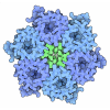
[English] 日本語
 Yorodumi
- PDB-1krc: CRYSTAL STRUCTURE OF KLEBSIELLA AEROGENES UREASE, ITS APOENZYME A... -
+ Open data
- Basic information
| Entry | Database: PDB / ID: 1krc | ||||||
|---|---|---|---|---|---|---|---|
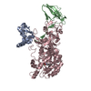
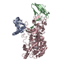
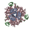
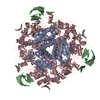
| Title | CRYSTAL STRUCTURE OF KLEBSIELLA AEROGENES UREASE, ITS APOENZYME AND TWO ACTIVE SITE MUTANTS | ||||||
 Components Components | (UREASE) x 3 | ||||||
 Keywords Keywords | HYDROLASE (UREA AMIDO) / ACTIVE SITE MUTANT / NICKEL METALLOENZYME | ||||||
| Function / homology |  Function and homology information Function and homology informationurease complex / urease / urease activity / urea catabolic process / nickel cation binding / cytoplasm Similarity search - Function | ||||||
| Biological species |  Klebsiella aerogenes (bacteria) Klebsiella aerogenes (bacteria) | ||||||
| Method |  X-RAY DIFFRACTION / Resolution: 2.5 Å X-RAY DIFFRACTION / Resolution: 2.5 Å | ||||||
 Authors Authors | Jabri, E. / Karplus, P.A. | ||||||
 Citation Citation |  Journal: Biochemistry / Year: 1996 Journal: Biochemistry / Year: 1996Title: Structures of the Klebsiella aerogenes urease apoenzyme and two active-site mutants. Authors: Jabri, E. / Karplus, P.A. #1:  Journal: Science / Year: 1995 Journal: Science / Year: 1995Title: The Crystal Structure of Urease from Klebsiella Aerogenes Authors: Jabri, E. / Carr, M.B. / Hausinger, R.P. / Karplus, P.A. #2:  Journal: Protein Sci. / Year: 1993 Journal: Protein Sci. / Year: 1993Title: Site-Directed Mutagenesis of Klebsiella Aerogenes Urease: Identification of Histidine Residues that Appear to Function in Nickel Ligation, Substrate Binding, and Catalysis Authors: Park, I.-L. / Hausinger, R.P. #3:  Journal: J.Mol.Biol. / Year: 1992 Journal: J.Mol.Biol. / Year: 1992Title: Preliminary Crystallographic Studies of Urease from Jack Bean and from Klebsiella Aerogenes Authors: Jabri, E. / Lee, M.H. / Hausinger, R.P. / Karplus, P.A. | ||||||
| History |
|
- Structure visualization
| Structure viewer | Molecule:  Molmil Molmil Jmol/JSmol Jmol/JSmol |
|---|
- Downloads & links
- Download
| PDBx/mmCIF format |  1krc.cif.gz 1krc.cif.gz | 156.7 KB | Display |  PDBx/mmCIF format PDBx/mmCIF format |
|---|---|---|---|---|
| PDB format |  pdb1krc.ent.gz pdb1krc.ent.gz | 122.5 KB | Display |  PDB format PDB format |
| PDBx/mmJSON format |  1krc.json.gz 1krc.json.gz | Tree view |  PDBx/mmJSON format PDBx/mmJSON format | |
| Others |  Other downloads Other downloads |
-Validation report
| Arichive directory |  https://data.pdbj.org/pub/pdb/validation_reports/kr/1krc https://data.pdbj.org/pub/pdb/validation_reports/kr/1krc ftp://data.pdbj.org/pub/pdb/validation_reports/kr/1krc ftp://data.pdbj.org/pub/pdb/validation_reports/kr/1krc | HTTPS FTP |
|---|
-Related structure data
- Links
Links
- Assembly
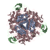
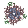
| Deposited unit | 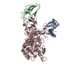
| ||||||||
|---|---|---|---|---|---|---|---|---|---|
| 1 | 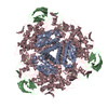
| ||||||||
| Unit cell |
| ||||||||
| Atom site foot note | 1: CIS PROLINE - PRO C 282 / 2: CIS PROLINE - PRO C 303 / 3: CIS PROLINE - PRO C 470 | ||||||||
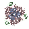
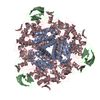
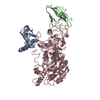
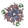
| Details | THREE NONIDENTICAL CHAINS, GAMMA (A), BETA (B), AND ALPHA (C) FORM ONE (ABC)-UNIT. THE ASYMMETRIC UNIT CONTAINS ONE (ABC)-UNIT. |
- Components
-Protein , 3 types, 3 molecules ABC
| #1: Protein | Mass: 11100.928 Da / Num. of mol.: 1 / Mutation: H(C 320)A / Source method: isolated from a natural source / Source: (natural)  Klebsiella aerogenes (bacteria) / Organ: BEAN / References: UniProt: P18316, urease Klebsiella aerogenes (bacteria) / Organ: BEAN / References: UniProt: P18316, urease |
|---|---|
| #2: Protein | Mass: 11712.239 Da / Num. of mol.: 1 / Mutation: H(C 320)A / Source method: isolated from a natural source / Source: (natural)  Klebsiella aerogenes (bacteria) / Organ: BEAN / References: UniProt: P18315, urease Klebsiella aerogenes (bacteria) / Organ: BEAN / References: UniProt: P18315, urease |
| #3: Protein | Mass: 60299.289 Da / Num. of mol.: 1 / Mutation: H(C 320)A / Source method: isolated from a natural source / Source: (natural)  Klebsiella aerogenes (bacteria) / Organ: BEAN / References: UniProt: P18314, urease Klebsiella aerogenes (bacteria) / Organ: BEAN / References: UniProt: P18314, urease |
-Non-polymers , 3 types, 162 molecules 


| #4: Chemical | | #5: Chemical | ChemComp-CO2 / | #6: Water | ChemComp-HOH / | |
|---|
-Details
| Nonpolymer details | HOH 1, THE CATALYTIC WATER, BRIDGES THE TWO NICKEL IONS. LYS 217 IS COVALENTLY MODIFIED AT NZ BY ...HOH 1, THE CATALYTIC WATER, BRIDGES THE TWO NICKEL IONS. LYS 217 IS COVALENTLY |
|---|
-Experimental details
-Experiment
| Experiment | Method:  X-RAY DIFFRACTION / Number of used crystals: 1 X-RAY DIFFRACTION / Number of used crystals: 1 |
|---|
- Sample preparation
| Crystal | Density Matthews: 2.5 Å3/Da / Density % sol: 50.73 % | ||||||||||||||||||||||||||||||||||||||||||||||||||||||
|---|---|---|---|---|---|---|---|---|---|---|---|---|---|---|---|---|---|---|---|---|---|---|---|---|---|---|---|---|---|---|---|---|---|---|---|---|---|---|---|---|---|---|---|---|---|---|---|---|---|---|---|---|---|---|---|
| Crystal grow | *PLUS Temperature: 25 ℃ / Method: vapor diffusion, hanging drop / pH: 7 / Details: Jabri, E., (1992) J.Mol.Biol., 227, 934. | ||||||||||||||||||||||||||||||||||||||||||||||||||||||
| Components of the solutions | *PLUS
|
-Data collection
| Diffraction source | Wavelength: 1.5418 Å |
|---|---|
| Detector | Type: XUONG-HAMLIN MULTIWIRE MARK II / Detector: AREA DETECTOR |
| Radiation | Monochromatic (M) / Laue (L): M / Scattering type: x-ray |
| Radiation wavelength | Wavelength: 1.5418 Å / Relative weight: 1 |
| Reflection | Resolution: 2.3→99.9 Å / Num. obs: 28672 / % possible obs: 99 % / Observed criterion σ(I): 0 / Redundancy: 4 % / Rmerge(I) obs: 0.094 |
| Reflection | *PLUS Highest resolution: 2.5 Å / Redundancy: 3.3 % / Rmerge(I) obs: 0.094 |
- Processing
| Software |
| ||||||||||||||||||||||||||||||||||||||||||||||||||||||||||||
|---|---|---|---|---|---|---|---|---|---|---|---|---|---|---|---|---|---|---|---|---|---|---|---|---|---|---|---|---|---|---|---|---|---|---|---|---|---|---|---|---|---|---|---|---|---|---|---|---|---|---|---|---|---|---|---|---|---|---|---|---|---|
| Refinement | Resolution: 2.5→10 Å / σ(F): 0 Details: RESIDUES 308 - 330 IN CHAIN C HAVE HIGH B VALUES. THEY CORRESPOND TO A MOBILE LOOP NEAR THE ACTIVE SITE.
| ||||||||||||||||||||||||||||||||||||||||||||||||||||||||||||
| Displacement parameters | Biso mean: 12.2 Å2 | ||||||||||||||||||||||||||||||||||||||||||||||||||||||||||||
| Refine analyze | Luzzati coordinate error obs: 0.25 Å | ||||||||||||||||||||||||||||||||||||||||||||||||||||||||||||
| Refinement step | Cycle: LAST / Resolution: 2.5→10 Å
| ||||||||||||||||||||||||||||||||||||||||||||||||||||||||||||
| Refine LS restraints |
| ||||||||||||||||||||||||||||||||||||||||||||||||||||||||||||
| Software | *PLUS Name:  X-PLOR / Version: 3.1 / Classification: refinement X-PLOR / Version: 3.1 / Classification: refinement | ||||||||||||||||||||||||||||||||||||||||||||||||||||||||||||
| Refinement | *PLUS Rfactor obs: 0.18 / Rfactor Rwork: 0.18 | ||||||||||||||||||||||||||||||||||||||||||||||||||||||||||||
| Solvent computation | *PLUS | ||||||||||||||||||||||||||||||||||||||||||||||||||||||||||||
| Displacement parameters | *PLUS | ||||||||||||||||||||||||||||||||||||||||||||||||||||||||||||
| Refine LS restraints | *PLUS Type: x_angle_deg / Dev ideal: 1.9 |
 Movie
Movie Controller


 PDBj
PDBj