[English] 日本語
Yorodumi
- PDB-1js8: Structure of a Functional Unit from Octopus Hemocyanin -

+
Open data


ID or keywords:

Loading...

-
Basic information

Entry
Database: PDB / ID: 1js8
TitleStructure of a Functional Unit from Octopus Hemocyanin
ComponentsHemocyanin
KeywordsOXYGEN STORAGE/TRANSPORT / GLYCOPROTEIN / MOLLUSC / OXYGEN-TRANSPORT / THIOETHER BOND / OXYGEN STORAGE-TRANSPORT COMPLEX
Function / homology
Function and homology information


oxygen carrier activity / oxidoreductase activity / metal ion binding
Similarity search - Function
Haemocyanin C-terminal domain / Hemocyanin; Chain: A, domain 2 / Haemocyanin beta-sandwich domain / Haemocyanin C-terminal domain superfamily / Haemocyanin beta-sandwich / di-copper center containing domain from catechol oxidase / Di-copper center containing domain from catechol oxidase / Tyrosinase CuA-binding region signature. / Common central domain of tyrosinase / Tyrosinase and hemocyanins CuB-binding region signature. ...Haemocyanin C-terminal domain / Hemocyanin; Chain: A, domain 2 / Haemocyanin beta-sandwich domain / Haemocyanin C-terminal domain superfamily / Haemocyanin beta-sandwich / di-copper center containing domain from catechol oxidase / Di-copper center containing domain from catechol oxidase / Tyrosinase CuA-binding region signature. / Common central domain of tyrosinase / Tyrosinase and hemocyanins CuB-binding region signature. / Tyrosinase copper-binding domain / Di-copper centre-containing domain superfamily / Sandwich / Orthogonal Bundle / Mainly Beta / Mainly Alpha
Similarity search - Domain/homology
CU2-O2 CLUSTER / alpha-D-mannopyranose / Hemocyanin G-type, units Oda to Odg
Similarity search - Component
Biological speciesOctopus dofleini (giant octopus)
MethodX-RAY DIFFRACTION / SYNCHROTRON / MIRAS, combined with partial MAD phasing / Resolution: 2.3 Å
AuthorsCuff, M.E. / Miller, K.I. / van Holde, K.E. / Hendrickson, W.A.
CitationJournal: J.Mol.Biol. / Year: 1998
Title: Crystal structure of a functional unit from Octopus hemocyanin.
Authors: Cuff, M.E. / Miller, K.I. / van Holde, K.E. / Hendrickson, W.A.
History
DepositionAug 16, 2001Deposition site: RCSB / Processing site: RCSB
Revision 1.0Oct 3, 2001Provider: repository / Type: Initial release
Revision 1.1Oct 16, 2007Group: Version format compliance
Revision 1.2Jul 13, 2011Group: Non-polymer description / Version format compliance
Revision 1.3Oct 4, 2017Group: Advisory / Refinement description / Category: pdbx_unobs_or_zero_occ_atoms / software
Item: _pdbx_unobs_or_zero_occ_atoms.label_asym_id / _software.name
Revision 2.0Jul 29, 2020Group: Advisory / Atomic model ...Advisory / Atomic model / Data collection / Derived calculations / Structure summary
Category: atom_site / chem_comp ...atom_site / chem_comp / entity / pdbx_branch_scheme / pdbx_chem_comp_identifier / pdbx_entity_branch / pdbx_entity_branch_descriptor / pdbx_entity_branch_link / pdbx_entity_branch_list / pdbx_entity_nonpoly / pdbx_nonpoly_scheme / pdbx_struct_assembly_gen / pdbx_struct_conn_angle / pdbx_unobs_or_zero_occ_atoms / pdbx_validate_close_contact / struct_asym / struct_conn / struct_conn_type / struct_site / struct_site_gen
Item: _atom_site.B_iso_or_equiv / _atom_site.Cartn_x ..._atom_site.B_iso_or_equiv / _atom_site.Cartn_x / _atom_site.Cartn_y / _atom_site.Cartn_z / _atom_site.auth_asym_id / _atom_site.auth_atom_id / _atom_site.auth_comp_id / _atom_site.auth_seq_id / _atom_site.label_asym_id / _atom_site.label_atom_id / _atom_site.label_comp_id / _atom_site.label_entity_id / _atom_site.type_symbol / _chem_comp.name / _chem_comp.type / _entity.formula_weight / _entity.pdbx_description / _entity.pdbx_number_of_molecules / _entity.type / _pdbx_struct_assembly_gen.asym_id_list / _pdbx_struct_conn_angle.ptnr1_label_asym_id / _pdbx_struct_conn_angle.ptnr2_label_asym_id / _pdbx_struct_conn_angle.ptnr3_label_asym_id / _pdbx_unobs_or_zero_occ_atoms.auth_asym_id / _pdbx_unobs_or_zero_occ_atoms.auth_atom_id / _pdbx_unobs_or_zero_occ_atoms.auth_seq_id / _pdbx_unobs_or_zero_occ_atoms.label_asym_id / _pdbx_unobs_or_zero_occ_atoms.label_atom_id / _pdbx_validate_close_contact.auth_asym_id_2 / _pdbx_validate_close_contact.auth_seq_id_2 / _struct_conn.conn_type_id / _struct_conn.id / _struct_conn.pdbx_dist_value / _struct_conn.pdbx_leaving_atom_flag / _struct_conn.pdbx_role / _struct_conn.ptnr1_auth_asym_id / _struct_conn.ptnr1_auth_comp_id / _struct_conn.ptnr1_auth_seq_id / _struct_conn.ptnr1_label_asym_id / _struct_conn.ptnr1_label_atom_id / _struct_conn.ptnr1_label_comp_id / _struct_conn.ptnr1_label_seq_id / _struct_conn.ptnr2_auth_asym_id / _struct_conn.ptnr2_auth_comp_id / _struct_conn.ptnr2_auth_seq_id / _struct_conn.ptnr2_label_asym_id / _struct_conn.ptnr2_label_atom_id / _struct_conn.ptnr2_label_comp_id / _struct_conn.ptnr2_label_seq_id / _struct_conn_type.id
Description: Carbohydrate remediation / Provider: repository / Type: Remediation
Remark 300
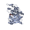
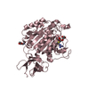
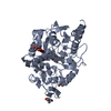
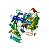
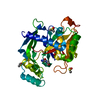
BIOMOLECULE: 1, 2 THIS ENTRY CONTAINS THE CRYSTALLOGRAPHIC ASYMMETRIC UNIT WHICH CONSISTS OF 2 ...BIOMOLECULE: 1, 2 THIS ENTRY CONTAINS THE CRYSTALLOGRAPHIC ASYMMETRIC UNIT WHICH CONSISTS OF 2 CHAIN(S). SEE REMARK 350 FOR INFORMATION ON GENERATING THE BIOLOGICAL MOLECULE(S). The native whole molecule cannot be generated from this information alone. Octopus hemocyanin is a 3.5MDa protein composed of 10 polypeptide subunits arranged about a D5 molecular symmetry axis. Each 350 kDa subunit contains 7 similar oxygen-binding functional units which occur serially along the polypeptide. Odg (this deposition) contains the C-terminal functional unit.

-
Structure visualization

Structure viewerMolecule:
MolmilJmol/JSmol

Downloads & links

-
Assembly

Deposited unit
A: Hemocyanin
B: Hemocyanin
hetero molecules


Theoretical massNumber of molelcules
Total (without water)92,2828
Polymers89,6202
Non-polymers2,6626
Water5,332296
1
A: Hemocyanin
hetero molecules


Theoretical massNumber of molelcules
Total (without water)46,3844
Polymers44,8101
Non-polymers1,5743
Water181
TypeNameSymmetry operationNumber
identity operation1_555x,y,z1
2
B: Hemocyanin
hetero molecules


Theoretical massNumber of molelcules
Total (without water)45,8984
Polymers44,8101
Non-polymers1,0883
Water181
TypeNameSymmetry operationNumber
identity operation1_555x,y,z1
Unit cell
Length a, b, c (Å)92.860, 168.390, 58.290
Angle α, β, γ (deg.)90.00, 90.00, 90.00
Int Tables number19
Space group name H-MP212121

-
Components

-
Protein , 1 types, 2 molecules AB

#1: Protein Hemocyanin / / HEMOCYANIN G-TYPE


Mass: 44810.062 Da / Num. of mol.: 2 / Fragment: C-terminal functional unit, Odg / Source method: isolated from a natural source
Details: C-TERMINAL PROTEOLYTIC FRAGMENT FROM 350KDA POLYPEPTIDE SUBUNIT. NATIVE MULTIMER IS 10 SUBUNITS EXHIBITING D5 SYMMETRY.
Source: (natural) Octopus dofleini (giant octopus) / Strain: G-type sequence / References: UniProt: O61363

-
Sugars , 4 types, 4 molecules

#2: Polysaccharide alpha-D-mannopyranose-(1-3)-[alpha-D-mannopyranose-(1-6)]alpha-D-mannopyranose-(1-6)-[beta-D- ...alpha-D-mannopyranose-(1-3)-[alpha-D-mannopyranose-(1-6)]alpha-D-mannopyranose-(1-6)-[beta-D-mannopyranose-(1-3)]beta-D-mannopyranose-(1-4)-2-acetamido-2-deoxy-beta-D-glucopyranose-(1-4)-2-acetamido-2-deoxy-alpha-D-glucopyranose


Type: oligosaccharide / Mass: 1235.105 Da / Num. of mol.: 1
Source method: isolated from a genetically manipulated source
DescriptorTypeProgram
DManpa1-3[DManpa1-6]DManpa1-6[DManpb1-3]DManpb1-4DGlcpNAcb1-4DGlcpNAca1-ROHGlycam Condensed SequenceGMML 1.0
WURCS=2.0/4,7,6/[a2122h-1a_1-5_2*NCC/3=O][a2122h-1b_1-5_2*NCC/3=O][a1122h-1b_1-5][a1122h-1a_1-5]/1-2-3-3-4-4-4/a4-b1_b4-c1_c3-d1_c6-e1_e3-f1_e6-g1WURCSPDB2Glycan 1.1.0
[][D-1-deoxy-GlcpNAc]{[(4+1)][b-D-GlcpNAc]{[(4+1)][b-D-Manp]{[(3+1)][b-D-Manp]{}[(6+1)][a-D-Manp]{[(3+1)][a-D-Manp]{}[(6+1)][a-D-Manp]{}}}}}LINUCSPDB-CARE
#3: Polysaccharide 2-acetamido-2-deoxy-beta-D-glucopyranose-(1-4)-2-acetamido-2-deoxy-beta-D-glucopyranose


Type: oligosaccharide / Mass: 424.401 Da / Num. of mol.: 1
Source method: isolated from a genetically manipulated source
DescriptorTypeProgram
DGlcpNAcb1-4DGlcpNAcb1-Glycam Condensed SequenceGMML 1.0
WURCS=2.0/1,2,1/[a2122h-1b_1-5_2*NCC/3=O]/1-1/a4-b1WURCSPDB2Glycan 1.1.0
[]{[(4+1)][b-D-GlcpNAc]{[(4+1)][b-D-GlcpNAc]{}}}LINUCSPDB-CARE
#4: Polysaccharide alpha-D-mannopyranose-(1-3)-[beta-D-mannopyranose-(1-6)]alpha-D-mannopyranose


Type: oligosaccharide / Mass: 504.438 Da / Num. of mol.: 1
Source method: isolated from a genetically manipulated source
DescriptorTypeProgram
DManpa1-3[DManpb1-6]DManpa1-ROHGlycam Condensed SequenceGMML 1.0
WURCS=2.0/2,3,2/[a1122h-1a_1-5][a1122h-1b_1-5]/1-1-2/a3-b1_a6-c1WURCSPDB2Glycan 1.1.0
[][D-1-deoxy-Manp]{[(3+1)][a-D-Manp]{}[(6+1)][b-D-Manp]{}}LINUCSPDB-CARE
#5: Sugar ChemComp-MAN / alpha-D-mannopyranose / Mannose


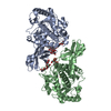
Type: D-saccharide, alpha linking / Mass: 180.156 Da / Num. of mol.: 1
Source method: isolated from a genetically manipulated source
Formula: C6H12O6
IdentifierTypeProgram
DManpaCONDENSED IUPAC CARBOHYDRATE SYMBOLGMML 1.0
a-D-mannopyranoseCOMMON NAMEGMML 1.0
a-D-ManpIUPAC CARBOHYDRATE SYMBOLPDB-CARE 1.0
ManSNFG CARBOHYDRATE SYMBOLGMML 1.0

-
Non-polymers , 2 types, 298 molecules

#6: Chemical ChemComp-CUO / CU2-O2 CLUSTER / CU-O2-CU LINKAGE


Mass: 159.091 Da / Num. of mol.: 2 / Source method: obtained synthetically / Formula: Cu2O2
#7: Water ChemComp-HOH / water / Water


Mass: 18.015 Da / Num. of mol.: 296 / Source method: isolated from a natural source / Formula: H2O

-
Details

Sequence detailsThe refinement statistics suggest that this well-ordered region contains a LEU.

-
Experimental details

-
Experiment

ExperimentMethod: X-RAY DIFFRACTION / Number of used crystals: 15

-
Sample preparation

CrystalDensity Matthews: 2.54 Å3/Da / Density % sol: 50 %
Crystal growTemperature: 277 K / Method: vapor diffusion, hanging drop / pH: 8.5
Details: PEG 3350, LITHIUM SULFATE, TRIS, pH 8.5, VAPOR DIFFUSION, HANGING DROP, temperature 277K
Crystal grow
*PLUS
Details: Cuff, M.E., (1990) J. Mol. Biol., 213, 11.

-
Data collection

Diffraction
IDMean temperature (K)Crystal-ID
12791
22981
Diffraction source
SourceSiteBeamlineTypeIDWavelength (Å)
SYNCHROTRONPhoton Factory PHOTON FACTORY BEAMLINE11.3796,1.3779,1.2000
SYNCHROTRONNSLS X4A20.98
Detector
TypeIDDetectorDate
FUJI1IMAGE PLATEJun 1, 1989
FUJI2IMAGE PLATEJun 1, 1994
Radiation
IDProtocolMonochromatic (M) / Laue (L)Scattering typeWavelength-IDMonochromator
1MADMx-ray1
2SINGLE WAVELENGTHMx-ray1SAGITALLY FOCUSED Si(111)
Radiation wavelength
IDWavelength (Å)Relative weight
11.37961
21.37791
31.21
40.981
Reflection
*PLUS
Highest resolution: 2.3 Å / % possible obs: 94.8 % / Rmerge(I) obs: 0.066

-
Processing

Software
NameVersionClassification
DENZOdata reduction
ROTAVATAdata reduction
MADSYSphasing
MLPHAREphasing
X-PLOR3.1refinement
CCP4(AGROVATAdata scaling
ROTAVATAdata scaling
RefinementMethod to determine structure: MIRAS, combined with partial MAD phasing
Resolution: 2.3→10 Å / σ(F): 2 / Stereochemistry target values: Engh & Huber
RfactorNum. reflectionSelection details
Rfree0.262 1614 RANDOM
Rwork0.202 --
obs0.201 38644 -
all-39114 -
Refinement stepCycle: LAST / Resolution: 2.3→10 Å
ProteinNucleic acidLigandSolventTotal
Num. atoms6134 0 163 296 6593
Refine LS restraints
Refine-IDTypeDev ideal
X-RAY DIFFRACTIONx_bond_d0.014
X-RAY DIFFRACTIONx_angle_d1.69
X-RAY DIFFRACTIONx_dihedral_angle_d24.29
X-RAY DIFFRACTIONx_improper_angle_d1.443
X-RAY DIFFRACTIONx_mcbond_it0.09
Refine LS restraints NCSWeight Biso : 1 / Weight position: 50
LS refinement shell
Resolution (Å)Rfactor RfreeNum. reflection RfreeRfactor RworkRefine-IDNum. reflection obs
2.3-2.380.2441490.214X-RAY DIFFRACTION2810
3.1-3.40.2442100.214X-RAY DIFFRACTION3723
4.79-100.2442080.214X-RAY DIFFRACTION3991
Software
*PLUS
Name: X-PLOR / Version: 3.1 / Classification: refinement
Refinement
*PLUS
Highest resolution: 2.3 Å / Lowest resolution: 10 Å / σ(F): 2
Solvent computation
*PLUS
Displacement parameters
*PLUS
Refine LS restraints
*PLUS
Refine-IDTypeDev ideal
X-RAY DIFFRACTIONx_angle_d
X-RAY DIFFRACTIONx_angle_deg1.69
X-RAY DIFFRACTIONx_dihedral_angle_d
X-RAY DIFFRACTIONx_dihedral_angle_deg24.29
X-RAY DIFFRACTIONx_improper_angle_d
X-RAY DIFFRACTIONx_improper_angle_deg1.443
LS refinement shell
*PLUS
Rfactor Rfree: 0.244 / Rfactor Rwork: 0.214

+
About Yorodumi

-
News

-
Feb 9, 2022. New format data for meta-information of EMDB entries

New format data for meta-information of EMDB entries

  • Version 3 of the EMDB header file is now the official format.
  • The previous official version 1.9 will be removed from the archive.

Related info.:EMDB header

External links:wwPDB to switch to version 3 of the EMDB data model

-
Aug 12, 2020. Covid-19 info

Covid-19 info

URL: https://pdbj.org/emnavi/covid19.php

New page: Covid-19 featured information page in EM Navigator.

Related info.:Covid-19 info / Mar 5, 2020. Novel coronavirus structure data

+
Mar 5, 2020. Novel coronavirus structure data

Novel coronavirus structure data

Related info.:Yorodumi Speices / Aug 12, 2020. Covid-19 info

External links:COVID-19 featured content - PDBj / Molecule of the Month (242):Coronavirus Proteases

+
Jan 31, 2019. EMDB accession codes are about to change! (news from PDBe EMDB page)

EMDB accession codes are about to change! (news from PDBe EMDB page)

  • The allocation of 4 digits for EMDB accession codes will soon come to an end. Whilst these codes will remain in use, new EMDB accession codes will include an additional digit and will expand incrementally as the available range of codes is exhausted. The current 4-digit format prefixed with “EMD-” (i.e. EMD-XXXX) will advance to a 5-digit format (i.e. EMD-XXXXX), and so on. It is currently estimated that the 4-digit codes will be depleted around Spring 2019, at which point the 5-digit format will come into force.
  • The EM Navigator/Yorodumi systems omit the EMD- prefix.

Related info.:Q: What is EMD? / ID/Accession-code notation in Yorodumi/EM Navigator

External links:EMDB Accession Codes are Changing Soon! / Contact to PDBj

+
Jul 12, 2017. Major update of PDB

Major update of PDB

  • wwPDB released updated PDB data conforming to the new PDBx/mmCIF dictionary.
  • This is a major update changing the version number from 4 to 5, and with Remediation, in which all the entries are updated.
  • In this update, many items about electron microscopy experimental information are reorganized (e.g. em_software).
  • Now, EM Navigator and Yorodumi are based on the updated data.

External links:wwPDB Remediation / Enriched Model Files Conforming to OneDep Data Standards Now Available in the PDB FTP Archive

-
Yorodumi

Thousand views of thousand structures

  • Yorodumi is a browser for structure data from EMDB, PDB, SASBDB, etc.
  • This page is also the successor to EM Navigator detail page, and also detail information page/front-end page for Omokage search.
  • The word "yorodu" (or yorozu) is an old Japanese word meaning "ten thousand". "mi" (miru) is to see.

Related info.:EMDB / PDB / SASBDB / Comparison of 3 databanks / Yorodumi Search / Aug 31, 2016. New EM Navigator & Yorodumi / Yorodumi Papers / Jmol/JSmol / Function and homology information / Changes in new EM Navigator and Yorodumi

Read more