 Movie
Movie Controller

[English] 日本語
 Yorodumi
- PDB-1jgm: High Resolution Structure of the Cadmium-containing Phosphotriest... -
+ Open data

- Basic information
| Entry | Database: PDB / ID: 1jgm | |||||||||
|---|---|---|---|---|---|---|---|---|---|---|
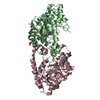
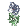
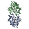
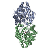
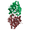
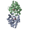
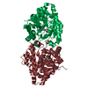
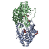
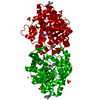
| Title | High Resolution Structure of the Cadmium-containing Phosphotriesterase from Pseudomonas diminuta | |||||||||
 Components Components | Phosphotriesterase | |||||||||
 Keywords Keywords | HYDROLASE / PTE / Cadmium | |||||||||
| Function / homology |  Function and homology information Function and homology informationaryldialkylphosphatase / aryldialkylphosphatase activity / zinc ion binding / plasma membrane Similarity search - Function | |||||||||
| Biological species |  Brevundimonas diminuta (bacteria) Brevundimonas diminuta (bacteria) | |||||||||
| Method |  X-RAY DIFFRACTION / SYNCHROTRON / FOURIER SYNTHESIS / Resolution: 1.3 Å X-RAY DIFFRACTION / SYNCHROTRON / FOURIER SYNTHESIS / Resolution: 1.3 Å | |||||||||
 Authors Authors | Benning, M.M. / Shim, H. / Raushel, F.M. / Holden, H.M. | |||||||||
 Citation Citation | Journal: Biochemistry / Year: 2001 Title: High resolution X-ray structures of different metal-substituted forms of phosphotriesterase from Pseudomonas diminuta. Authors: Benning, M.M. / Shim, H. / Raushel, F.M. / Holden, H.M. | |||||||||
| History |
|
- Structure visualization
| Structure viewer | Molecule: MolmilJmol/JSmol |
|---|
- Downloads & links
-Download
| PDBx/mmCIF format | 1jgm.cif.gz | 165.8 KB | Display | PDBx/mmCIF format |
|---|---|---|---|---|
| PDB format | pdb1jgm.ent.gz | 127 KB | Display | PDB format |
| PDBx/mmJSON format | 1jgm.json.gz | Tree view | PDBx/mmJSON format | |
| Others |  Other downloads Other downloads |
-Validation report
| Arichive directory | https://data.pdbj.org/pub/pdb/validation_reports/jg/1jgmftp://data.pdbj.org/pub/pdb/validation_reports/jg/1jgm | HTTPS FTP |
|---|
-Related structure data


-Links
 PDBj
PDBj


- Assembly
| Deposited unit | 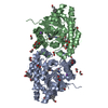
| ||||||||
|---|---|---|---|---|---|---|---|---|---|
| 1 |
| ||||||||
| Unit cell |
| ||||||||
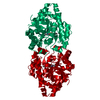
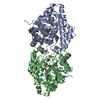
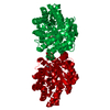
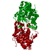
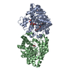
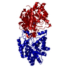
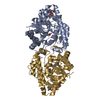
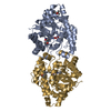
| Details | The biological asembly is a dimer that is described by chains A & B in the asymmetric unit |
-Components
-Protein , 1 types, 2 molecules AB
| #1: Protein | Mass: 36375.402 Da / Num. of mol.: 2 Source method: isolated from a genetically manipulated source Source: (gene. exp.) Brevundimonas diminuta (bacteria) / Gene: OPD / Production host: |
|---|
-Non-polymers , 5 types, 776 molecules 


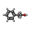

| #2: Chemical | ChemComp-CD /  Mass: 112.411 Da / Num. of mol.: 4 / Source method: obtained synthetically / Formula: Cd Mass: 112.411 Da / Num. of mol.: 4 / Source method: obtained synthetically / Formula: Cd#3: Chemical |  Mass: 22.990 Da / Num. of mol.: 2 / Source method: obtained synthetically / Formula: Na Mass: 22.990 Da / Num. of mol.: 2 / Source method: obtained synthetically / Formula: Na#4: Chemical | ChemComp-EDO /  Mass: 62.068 Da / Num. of mol.: 27 / Source method: obtained synthetically / Formula: C2H6O2 Mass: 62.068 Da / Num. of mol.: 27 / Source method: obtained synthetically / Formula: C2H6O2#5: Chemical |  Mass: 122.164 Da / Num. of mol.: 3 / Source method: obtained synthetically / Formula: C8H10O Mass: 122.164 Da / Num. of mol.: 3 / Source method: obtained synthetically / Formula: C8H10O#6: Water | ChemComp-HOH / | Mass: 18.015 Da / Num. of mol.: 740 / Source method: isolated from a natural source / Formula: H2O |
|---|
-Details
| Has protein modification | Y |
|---|
-Experimental details
-Experiment
| Experiment | Method: X-RAY DIFFRACTION / Number of used crystals: 1 |
|---|
- Sample preparation
| Crystal | Density Matthews: 2.72 Å3/Da / Density % sol: 54.7 % | ||||||||||||||||||||||||||||||||||||
|---|---|---|---|---|---|---|---|---|---|---|---|---|---|---|---|---|---|---|---|---|---|---|---|---|---|---|---|---|---|---|---|---|---|---|---|---|---|
| Crystal grow | Temperature: 277 K / Method: batch/microseeding / pH: 9 Details: PEG-8000, sodium chloride, phenylethyl alcohol, diethyl-4-methylbenzylphosphate, CHES, pH 9, Batch/microseeding, temperature 277K | ||||||||||||||||||||||||||||||||||||
| Crystal grow | *PLUS Temperature: 4 ℃ / pH: 9 / Method: vapor diffusion, hanging drop / Details: Benning, M.M., (1995) Biochemistry, 34, 7973. | ||||||||||||||||||||||||||||||||||||
| Components of the solutions | *PLUS
|
-Data collection
| Diffraction | Mean temperature: 100 K |
|---|---|
| Diffraction source | Source: SYNCHROTRON / Site: APS  / Beamline: 19-ID / Wavelength: 0.71 Å / Beamline: 19-ID / Wavelength: 0.71 Å |
| Detector | Type: SBC-2 / Detector: CCD / Date: Dec 17, 1999 |
| Radiation | Monochromator: SI 111 / Protocol: SINGLE WAVELENGTH / Monochromatic (M) / Laue (L): M / Scattering type: x-ray |
| Radiation wavelength | Wavelength: 0.71 Å / Relative weight: 1 |
| Reflection | Resolution: 1.3→30 Å / Num. all: 187426 / Num. obs: 187426 / % possible obs: 98 % / Observed criterion σ(F): 0 / Observed criterion σ(I): 0 / Redundancy: 3.4 % / Rmerge(I) obs: 0.069 / Net I/σ(I): 32 |
| Reflection shell | Resolution: 1.3→1.37 Å / Redundancy: 3.2 % / Rmerge(I) obs: 0.184 / % possible all: 99 |
| Reflection | *PLUS Num. measured all: 1231444 |
| Reflection shell | *PLUS % possible obs: 99 % |
- Processing
| Software |
| |||||||||||||||||||||||||
|---|---|---|---|---|---|---|---|---|---|---|---|---|---|---|---|---|---|---|---|---|---|---|---|---|---|---|
| Refinement | Method to determine structure: FOURIER SYNTHESIS / Resolution: 1.3→30 Å / σ(F): 0 / σ(I): 0 / Stereochemistry target values: Engh & Huber
| |||||||||||||||||||||||||
| Refinement step | Cycle: LAST / Resolution: 1.3→30 Å
| |||||||||||||||||||||||||
| Refine LS restraints |
| |||||||||||||||||||||||||
| Software | *PLUS Name: TNT / Classification: refinement | |||||||||||||||||||||||||
| Refinement | *PLUS σ(F): 0 / % reflection Rfree: 4.9 % / Rfactor all: 0.204 / Rfactor Rwork: 0.202 | |||||||||||||||||||||||||
| Solvent computation | *PLUS | |||||||||||||||||||||||||
| Displacement parameters | *PLUS | |||||||||||||||||||||||||
| Refine LS restraints | *PLUS Type: t_angle_deg / Dev ideal: 2.1 |
