 Movie
Movie Controller

[English] 日本語
 Yorodumi
- PDB-1iuu: P-HYDROXYBENZOATE HYDROXYLASE COMPLEXED WITH 4-AMINOBENZOATE AT PH 9.4 -
+ Open data

- Basic information
| Entry | Database: PDB / ID: 1iuu | ||||||
|---|---|---|---|---|---|---|---|
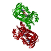
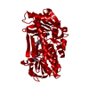
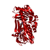
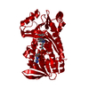
| Title | P-HYDROXYBENZOATE HYDROXYLASE COMPLEXED WITH 4-AMINOBENZOATE AT PH 9.4 | ||||||
 Components Components | P-HYDROXYBENZOATE HYDROXYLASE | ||||||
 Keywords Keywords | OXIDOREDUCTASE | ||||||
| Function / homology |  Function and homology information Function and homology information4-hydroxybenzoate 3-monooxygenase (NADPH) activity / 4-hydroxybenzoate 3-monooxygenase / 4-hydroxybenzoate 3-monooxygenase activity / : / FAD binding / flavin adenine dinucleotide binding / oxidoreductase activity Similarity search - Function | ||||||
| Biological species |   Pseudomonas aeruginosa (bacteria) Pseudomonas aeruginosa (bacteria) | ||||||
| Method |  X-RAY DIFFRACTION / Resolution: 2 Å X-RAY DIFFRACTION / Resolution: 2 Å | ||||||
 Authors Authors | Gatti, D.L. / Entsch, B. / Ballou, D.P. / Ludwig, M.L. | ||||||
 Citation Citation | Journal: Biochemistry / Year: 1996 Title: pH-dependent structural changes in the active site of p-hydroxybenzoate hydroxylase point to the importance of proton and water movements during catalysis. Authors: Gatti, D.L. / Entsch, B. / Ballou, D.P. / Ludwig, M.L. #1: Journal: Biochemistry / Year: 1994Title: Crystal Structures of Wild-Type P-Hydroxybenzoate Hydroxylase Complexed with 4-Aminobenzoate, 2,4-Dihydrobenzoate and 2-Hydroxy-4-Aminobenzoate and of the Tyr222Ala Mutant Complexed with 2- ...Title: Crystal Structures of Wild-Type P-Hydroxybenzoate Hydroxylase Complexed with 4-Aminobenzoate, 2,4-Dihydrobenzoate and 2-Hydroxy-4-Aminobenzoate and of the Tyr222Ala Mutant Complexed with 2-Hydroxy-4-Aminobenzoate.Evidence for a Proton Channel and a New Binding Mode of the Flavin Ring Authors: Schreuder, H.A. / Mattevi, A. / Obmolova, G. / Kalk, K.H. / Hol, W.G.J. #2: Journal: Biochemistry / Year: 1994Title: Crystal Structures of Mutant Pseudomonas Aeruginosa P-Hydroxybenzoate Hydroxylases:The Tyr201Phe, Tyr385Phe and Asn300Asp Variants Authors: Lah, M.S. / Palfey, B.A. / Schreuder, H.A. / Ludwig, M.L. #3: Journal: J.Biol.Chem. / Year: 1991Title: Catalytic Function of Tyrosine Residues in Para-Hydroxybenzoate Hydroxylase as Determined by the Study of Site-Directed Mutants Authors: Entsch, B. / Palfey, B.A. / Ballou, D.P. / Massey, V. | ||||||
| History |
|
- Structure visualization
| Structure viewer | Molecule: MolmilJmol/JSmol |
|---|
- Downloads & links
-Download
| PDBx/mmCIF format | 1iuu.cif.gz | 98 KB | Display | PDBx/mmCIF format |
|---|---|---|---|---|
| PDB format | pdb1iuu.ent.gz | 74 KB | Display | PDB format |
| PDBx/mmJSON format | 1iuu.json.gz | Tree view | PDBx/mmJSON format | |
| Others |  Other downloads Other downloads |
-Validation report
| Arichive directory | https://data.pdbj.org/pub/pdb/validation_reports/iu/1iuuftp://data.pdbj.org/pub/pdb/validation_reports/iu/1iuu | HTTPS FTP |
|---|
-Related structure data


-Links
 PDBj
PDBj- Assembly
| Deposited unit | 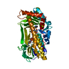
| ||||||||
|---|---|---|---|---|---|---|---|---|---|
| 1 | 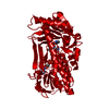
| ||||||||
| Unit cell |
| ||||||||
| Atom site foot note | 1: CIS PROLINE - PRO 275 |
-Components
| #1: Protein | Mass: 44382.547 Da / Num. of mol.: 1 Source method: isolated from a genetically manipulated source Details: PH 9.4 STRUCTURE / Source: (gene. exp.) Pseudomonas aeruginosa (bacteria)References: UniProt: P20586, 4-hydroxybenzoate 3-monooxygenase |
|---|---|
| #2: Chemical | ChemComp-FAD /   Mass: 785.550 Da / Num. of mol.: 1 / Source method: obtained synthetically / Formula: C27H33N9O15P2 / Comment: FAD*YM Mass: 785.550 Da / Num. of mol.: 1 / Source method: obtained synthetically / Formula: C27H33N9O15P2 / Comment: FAD*YM |
| #3: Chemical | ChemComp-PAB /   Mass: 137.136 Da / Num. of mol.: 1 / Source method: obtained synthetically / Formula: C7H7NO2 Mass: 137.136 Da / Num. of mol.: 1 / Source method: obtained synthetically / Formula: C7H7NO2 |
| #4: Water | ChemComp-HOH /  Mass: 18.015 Da / Num. of mol.: 218 / Source method: isolated from a natural source / Formula: H2O Mass: 18.015 Da / Num. of mol.: 218 / Source method: isolated from a natural source / Formula: H2O |
-Experimental details
-Experiment
| Experiment | Method: X-RAY DIFFRACTION / Number of used crystals: 1 |
|---|
- Sample preparation
| Crystal | Density Matthews: 2.61 Å3/Da / Density % sol: 52.84 % | ||||||||||||||||||||||||||||||||||||||||||||||||
|---|---|---|---|---|---|---|---|---|---|---|---|---|---|---|---|---|---|---|---|---|---|---|---|---|---|---|---|---|---|---|---|---|---|---|---|---|---|---|---|---|---|---|---|---|---|---|---|---|---|
| Crystal grow | pH: 9.4 / Details: pH 9.4 | ||||||||||||||||||||||||||||||||||||||||||||||||
| Crystal grow | *PLUS Temperature: 4-23 ℃ / pH: 7.4 / Method: interface diffusion | ||||||||||||||||||||||||||||||||||||||||||||||||
| Components of the solutions | *PLUS
|
-Data collection
| Diffraction | Mean temperature: 295 K |
|---|---|
| Diffraction source | Wavelength: 1.5418 Å |
| Detector | Type: SDMS / Detector: AREA DETECTOR / Details: 0.5 MM COLLIMATOR |
| Radiation | Monochromator: GRAPHITE / Monochromatic (M) / Laue (L): M / Scattering type: x-ray |
| Radiation wavelength | Wavelength: 1.5418 Å / Relative weight: 1 |
| Reflection | Resolution: 2→15 Å / % possible obs: 98.73 % / Redundancy: 5.96 % / Rmerge(I) obs: 0.112 / Net I/σ(I): 8.91 |
| Reflection shell | Resolution: 2→2.15 Å / Rmerge(I) obs: 0.336 / Mean I/σ(I) obs: 2.08 / % possible all: 95.34 |
| Reflection | *PLUS Highest resolution: 2 Å / Lowest resolution: 15 Å / Num. obs: 31276 / % possible obs: 98.73 % / Redundancy: 8.25 % |
- Processing
| Software |
| ||||||||||||||||||||||||||||||||||||||||||||||||||||||||||||
|---|---|---|---|---|---|---|---|---|---|---|---|---|---|---|---|---|---|---|---|---|---|---|---|---|---|---|---|---|---|---|---|---|---|---|---|---|---|---|---|---|---|---|---|---|---|---|---|---|---|---|---|---|---|---|---|---|---|---|---|---|---|
| Refinement | Resolution: 2→15 Å / σ(F): 0 Details: THE HYDROGEN BOND DISTANCE CUTOFF USED FOR THE TURNS IS 3.5 ANGSTROMS. THE CA(X) TO CA(X+4) DISTANCE IS LESS THAN 6.0 ANGSTROMS. THE ANGLES ARE FROM ROBSON AND GARNIER, (1986), 'INTRODUCTION ...Details: THE HYDROGEN BOND DISTANCE CUTOFF USED FOR THE TURNS IS 3.5 ANGSTROMS. THE CA(X) TO CA(X+4) DISTANCE IS LESS THAN 6.0 ANGSTROMS. THE ANGLES ARE FROM ROBSON AND GARNIER, (1986), 'INTRODUCTION TO PROTEINS AND PROTEIN ENGINEERING', ELSEVIER, AMSTERDAM. TYPE PHI2 PSI2 PHI3 PSI3 I -75(+-65) -30(+-40) -90(+-40) -15 TO 40 II -60(+-40) 120(+-40) 90(+-40) 0(+-40) III -75(+-65) -30(+-40) -60(+-40) -15 TO -70 I(PRIME) 75(+-65) 30(+-40) 90(+-40) -40 TO 15 II(PRIME) 60(+-40) -120(+-40) -90(+-40) 0(+-40) III(PRIME) 75(+-65) 30(+-40) 60(+-40) 15 TO 70 MODIFIED CYSTEINE WITH ADDITIONAL ELECTRON DENSITY NEAR THE SULFUR AT X=5.26, Y=107.89, Z=71.02 - CYS 116. THE TYPE OF CHEMICAL MODIFICATION CANNOT BE UNAMBIGUOUSLY DETERMINED FROM THE ELECTRON DENSITY THE SIDE CHAINS OF RESIDUES 135, 136, 146, 392, 393, 394 ARE NOT ORDERED. THE MODEL IS DERIVED FROM A COMBINATION OF FIT TO RESIDUAL DENSITY AND ENERGY MINIMIZATION.
| ||||||||||||||||||||||||||||||||||||||||||||||||||||||||||||
| Refinement step | Cycle: LAST / Resolution: 2→15 Å
| ||||||||||||||||||||||||||||||||||||||||||||||||||||||||||||
| Refine LS restraints |
| ||||||||||||||||||||||||||||||||||||||||||||||||||||||||||||
| Software | *PLUS Name: X-PLOR / Classification: refinement | ||||||||||||||||||||||||||||||||||||||||||||||||||||||||||||
| Refine LS restraints | *PLUS
|
