 Movie
Movie Controller

[English] 日本語
 Yorodumi
- PDB-1i1l: CRYSTAL STRUCTURE OF ESCHELICHIA COLI BRANCHED-CHAIN AMINO ACID A... -
+ Open data

- Basic information
| Entry | Database: PDB / ID: 1i1l | ||||||
|---|---|---|---|---|---|---|---|
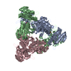
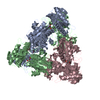
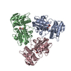
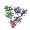
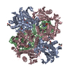
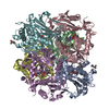
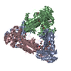
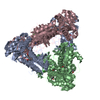
| Title | CRYSTAL STRUCTURE OF ESCHELICHIA COLI BRANCHED-CHAIN AMINO ACID AMINOTRANSFERASE. | ||||||
 Components Components | BRANCHED-CHAIN AMINO ACID AMINOTRANSFERASE | ||||||
 Keywords Keywords | TRANSFERASE / AMINOTRANSFERASE / HEXAMER / PLP / 2-METHYLLEUCINE | ||||||
| Function / homology |  Function and homology information Function and homology informationL-aspartate biosynthetic process / branched-chain-amino-acid:2-oxoglutarate transaminase activity / L-valine:2-oxoglutarate transaminase activity / L-isoleucine:2-oxoglutarate transaminase activity / L-leucine:2-oxoglutarate transaminase activity / branched-chain amino acid biosynthetic process / branched-chain-amino-acid transaminase / L-leucine biosynthetic process / L-valine biosynthetic process / : ...L-aspartate biosynthetic process / branched-chain-amino-acid:2-oxoglutarate transaminase activity / L-valine:2-oxoglutarate transaminase activity / L-isoleucine:2-oxoglutarate transaminase activity / L-leucine:2-oxoglutarate transaminase activity / branched-chain amino acid biosynthetic process / branched-chain-amino-acid transaminase / L-leucine biosynthetic process / L-valine biosynthetic process / : / identical protein binding / cytosol Similarity search - Function | ||||||
| Biological species |  | ||||||
| Method |  X-RAY DIFFRACTION / MOLECULAR REPLACEMENT / Resolution: 2.4 Å X-RAY DIFFRACTION / MOLECULAR REPLACEMENT / Resolution: 2.4 Å | ||||||
 Authors Authors | Okada, K. / Hirotsu, K. / Hayashi, H. / Kagamiyama, H. | ||||||
 Citation Citation | Journal: Biochemistry / Year: 2001 Title: Structures of Escherichia coli branched-chain amino acid aminotransferase and its complexes with 4-methylvalerate and 2-methylleucine: induced fit and substrate recognition of the enzyme. Authors: Okada, K. / Hirotsu, K. / Hayashi, H. / Kagamiyama, H. | ||||||
| History |
|
- Structure visualization
| Structure viewer | Molecule: MolmilJmol/JSmol |
|---|
- Downloads & links
-Download
| PDBx/mmCIF format | 1i1l.cif.gz | 192.3 KB | Display | PDBx/mmCIF format |
|---|---|---|---|---|
| PDB format | pdb1i1l.ent.gz | 152.5 KB | Display | PDB format |
| PDBx/mmJSON format | 1i1l.json.gz | Tree view | PDBx/mmJSON format | |
| Others |  Other downloads Other downloads |
-Validation report
| Arichive directory | https://data.pdbj.org/pub/pdb/validation_reports/i1/1i1lftp://data.pdbj.org/pub/pdb/validation_reports/i1/1i1l | HTTPS FTP |
|---|
-Related structure data


-Links
 PDBj
PDBj
- Assembly
| Deposited unit | 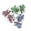
| ||||||||
|---|---|---|---|---|---|---|---|---|---|
| 1 | 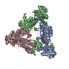
| ||||||||
| 2 | 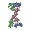
| ||||||||
| Unit cell |
|
-Components
| #1: Protein | Mass: 34131.586 Da / Num. of mol.: 3 Source method: isolated from a genetically manipulated source Source: (gene. exp.) References: UniProt: P00510, UniProt: P0AB80*PLUS, branched-chain-amino-acid transaminase #2: Chemical |   Mass: 247.142 Da / Num. of mol.: 3 / Source method: obtained synthetically / Formula: C8H10NO6P Mass: 247.142 Da / Num. of mol.: 3 / Source method: obtained synthetically / Formula: C8H10NO6P#3: Chemical |   Type: L-peptide linking / Mass: 145.199 Da / Num. of mol.: 3 / Source method: obtained synthetically / Formula: C7H15NO2 Type: L-peptide linking / Mass: 145.199 Da / Num. of mol.: 3 / Source method: obtained synthetically / Formula: C7H15NO2#4: Water | ChemComp-HOH / |  Mass: 18.015 Da / Num. of mol.: 304 / Source method: isolated from a natural source / Formula: H2O Mass: 18.015 Da / Num. of mol.: 304 / Source method: isolated from a natural source / Formula: H2O |
|---|
-Experimental details
-Experiment
| Experiment | Method: X-RAY DIFFRACTION / Number of used crystals: 1 |
|---|
- Sample preparation
| Crystal | Density Matthews: 2.72 Å3/Da / Density % sol: 54.73 % | ||||||||||||||||||||||||||||||||||||||||||
|---|---|---|---|---|---|---|---|---|---|---|---|---|---|---|---|---|---|---|---|---|---|---|---|---|---|---|---|---|---|---|---|---|---|---|---|---|---|---|---|---|---|---|---|
| Crystal grow | Temperature: 293 K / Method: vapor diffusion, hanging drop / pH: 7.5 Details: PEG400, pH 7.5, VAPOR DIFFUSION, HANGING DROP, temperature 293K | ||||||||||||||||||||||||||||||||||||||||||
| Crystal grow | *PLUS Temperature: 20 ℃ | ||||||||||||||||||||||||||||||||||||||||||
| Components of the solutions | *PLUS
|
-Data collection
| Diffraction | Mean temperature: 293 K |
|---|---|
| Diffraction source | Source: ROTATING ANODE / Type: RIGAKU RU200H / Wavelength: 1.5418 Å |
| Detector | Type: RIGAKU RAXIS IIC / Detector: IMAGE PLATE / Date: Jan 15, 1997 |
| Radiation | Monochromator: GRAPHITE / Protocol: SINGLE WAVELENGTH / Monochromatic (M) / Laue (L): M / Scattering type: x-ray |
| Radiation wavelength | Wavelength: 1.5418 Å / Relative weight: 1 |
| Reflection | Resolution: 2.4→40 Å / Observed criterion σ(F): 0 / Observed criterion σ(I): 0 |
| Reflection | *PLUS Lowest resolution: 40 Å / Num. obs: 42170 / % possible obs: 98.6 % / Num. measured all: 132437 / Rmerge(I) obs: 0.057 |
| Reflection shell | *PLUS Highest resolution: 2.4 Å / Lowest resolution: 2.51 Å / % possible obs: 98.4 % / Rmerge(I) obs: 0.181 |
- Processing
| Software |
| |||||||||||||||
|---|---|---|---|---|---|---|---|---|---|---|---|---|---|---|---|---|
| Refinement | Method to determine structure: MOLECULAR REPLACEMENT / Resolution: 2.4→40 Å / σ(F): 0 / σ(I): 0 / Stereochemistry target values: Engh & Huber /
| |||||||||||||||
| Refinement step | Cycle: LAST / Resolution: 2.4→40 Å
| |||||||||||||||
| Refine LS restraints |
| |||||||||||||||
| Software | *PLUS Name: X-PLOR / Version: 3.851 / Classification: refinement | |||||||||||||||
| Refinement | *PLUS Highest resolution: 2.4 Å / Lowest resolution: 40 Å / σ(F): 0 / Rfactor obs: 0.182 | |||||||||||||||
| Solvent computation | *PLUS | |||||||||||||||
| Displacement parameters | *PLUS | |||||||||||||||
| Refine LS restraints | *PLUS Type: x_angle_deg / Dev ideal: 1.3 | |||||||||||||||
| LS refinement shell | *PLUS Rfactor Rfree: 0.283 / Rfactor obs: 0.25 |
