 Movie
Movie Controller

[English] 日本語
 Yorodumi
- PDB-1hqs: CRYSTAL STRUCTURE OF ISOCITRATE DEHYDROGENASE FROM BACILLUS SUBTILIS -
+ Open data

- Basic information
| Entry | Database: PDB / ID: 1hqs | ||||||
|---|---|---|---|---|---|---|---|
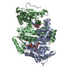
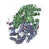
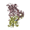
| Title | CRYSTAL STRUCTURE OF ISOCITRATE DEHYDROGENASE FROM BACILLUS SUBTILIS | ||||||
 Components Components | ISOCITRATE DEHYDROGENASE | ||||||
 Keywords Keywords | OXIDOREDUCTASE / glyoxylate bypass / BsIDH / tricarboxylic acid cycle / protein phosphorylation / NADP | ||||||
| Function / homology |  Function and homology information Function and homology informationisocitrate dehydrogenase (NADP+) / isocitrate dehydrogenase (NADP+) activity / glyoxylate cycle / tricarboxylic acid cycle / NAD binding / magnesium ion binding Similarity search - Function | ||||||
| Biological species |  | ||||||
| Method |  X-RAY DIFFRACTION / SYNCHROTRON / MOLECULAR REPLACEMENT / Resolution: 1.55 Å X-RAY DIFFRACTION / SYNCHROTRON / MOLECULAR REPLACEMENT / Resolution: 1.55 Å | ||||||
 Authors Authors | Singh, S.K. / Matsuno, K. / LaPorte, D.C. / Banaszak, L.J. | ||||||
 Citation Citation | Journal: J.Biol.Chem. / Year: 2001 Title: Crystal structure of Bacillus subtilis isocitrate dehydrogenase at 1.55 A. Insights into the nature of substrate specificity exhibited by Escherichia coli isocitrate dehydrogenase kinase/phosphatase. Authors: Singh, S.K. / Matsuno, K. / LaPorte, D.C. / Banaszak, L.J. | ||||||
| History |
| ||||||
| Remark 600 | HETEROGEN Cys118 from both monomers have been modified with beta-mercaptoethanol. |
- Structure visualization
| Structure viewer | Molecule: MolmilJmol/JSmol |
|---|
- Downloads & links
-Download
| PDBx/mmCIF format | 1hqs.cif.gz | 192.5 KB | Display | PDBx/mmCIF format |
|---|---|---|---|---|
| PDB format | pdb1hqs.ent.gz | 152.2 KB | Display | PDB format |
| PDBx/mmJSON format | 1hqs.json.gz | Tree view | PDBx/mmJSON format | |
| Others |  Other downloads Other downloads |
-Validation report
| Arichive directory | https://data.pdbj.org/pub/pdb/validation_reports/hq/1hqsftp://data.pdbj.org/pub/pdb/validation_reports/hq/1hqs | HTTPS FTP |
|---|
-Related structure data
| Related structure data | 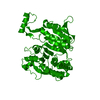 3icdS S: Starting model for refinement |
|---|---|
| Similar structure data |


-Links
 PDBj
PDBj

- Assembly
| Deposited unit | 
| ||||||||
|---|---|---|---|---|---|---|---|---|---|
| 1 |
| ||||||||
| Unit cell |
|
-Components
| #1: Protein | Mass: 46544.668 Da / Num. of mol.: 2 Source method: isolated from a genetically manipulated source Source: (gene. exp.) References: UniProt: P39126, isocitrate dehydrogenase (NADP+) #2: Chemical |   Mass: 192.124 Da / Num. of mol.: 2 / Source method: obtained synthetically / Formula: C6H8O7 Mass: 192.124 Da / Num. of mol.: 2 / Source method: obtained synthetically / Formula: C6H8O7#3: Chemical | ChemComp-PGO /   Mass: 76.094 Da / Num. of mol.: 5 / Source method: obtained synthetically / Formula: C3H8O2 Mass: 76.094 Da / Num. of mol.: 5 / Source method: obtained synthetically / Formula: C3H8O2#4: Chemical |   Mass: 76.094 Da / Num. of mol.: 2 / Source method: obtained synthetically / Formula: C3H8O2 Mass: 76.094 Da / Num. of mol.: 2 / Source method: obtained synthetically / Formula: C3H8O2#5: Water | ChemComp-HOH / |  Mass: 18.015 Da / Num. of mol.: 608 / Source method: isolated from a natural source / Formula: H2O Mass: 18.015 Da / Num. of mol.: 608 / Source method: isolated from a natural source / Formula: H2OHas protein modification | Y | |
|---|
-Experimental details
-Experiment
| Experiment | Method: X-RAY DIFFRACTION / Number of used crystals: 1 |
|---|
- Sample preparation
| Crystal | Density Matthews: 2.5 Å3/Da / Density % sol: 44.38 % | ||||||||||||||||||||||||||||||
|---|---|---|---|---|---|---|---|---|---|---|---|---|---|---|---|---|---|---|---|---|---|---|---|---|---|---|---|---|---|---|---|
| Crystal grow | Temperature: 291 K / Method: vapor diffusion, hanging drop / pH: 4.9 Details: 23% PEG 4000, 18% propylene glycol, 0.1 M citrate, pH 4.9, VAPOR DIFFUSION, HANGING DROP, temperature 291K | ||||||||||||||||||||||||||||||
| Crystal grow | *PLUS Temperature: 18 ℃ | ||||||||||||||||||||||||||||||
| Components of the solutions | *PLUS
|
-Data collection
| Diffraction | Mean temperature: 110 K |
|---|---|
| Diffraction source | Source: SYNCHROTRON / Site: APS  / Beamline: 19-ID / Wavelength: 1.0332 Å / Beamline: 19-ID / Wavelength: 1.0332 Å |
| Detector | Type: APS-1 / Detector: CCD / Date: Feb 6, 1998 / Details: vertically focusing mirror |
| Radiation | Monochromator: Sagitally focusing crystal/double crystal monochromator Si-111 Protocol: SINGLE WAVELENGTH / Monochromatic (M) / Laue (L): M / Scattering type: x-ray |
| Radiation wavelength | Wavelength: 1.0332 Å / Relative weight: 1 |
| Reflection | Resolution: 1.5→99 Å / Num. all: 244628 / Num. obs: 114797 / % possible obs: 88.5 % / Observed criterion σ(F): 0 / Observed criterion σ(I): 0 / Redundancy: 2.13 % / Biso Wilson estimate: 20.5 Å2 / Rmerge(I) obs: 0.077 / Net I/σ(I): 11 |
| Reflection shell | Resolution: 1.5→1.55 Å / Redundancy: 2.67 % / Rmerge(I) obs: 0.321 / Num. unique all: 6004 / % possible all: 46.6 |
| Reflection | *PLUS Highest resolution: 1.5 Å / Num. measured all: 244628 |
- Processing
| Software |
| ||||||||||||||||||||||||||||||||||||||||
|---|---|---|---|---|---|---|---|---|---|---|---|---|---|---|---|---|---|---|---|---|---|---|---|---|---|---|---|---|---|---|---|---|---|---|---|---|---|---|---|---|---|
| Refinement | Method to determine structure: MOLECULAR REPLACEMENT Starting model: PDB ENTRY 3ICD Resolution: 1.55→20 Å / Rfactor Rfree error: 0.003 / Data cutoff high absF: 10000000 / Data cutoff low absF: 0 / Isotropic thermal model: Restrained / Cross valid method: THROUGHOUT / σ(F): 0 / σ(I): 0 / Stereochemistry target values: Engh & Huber Details: Used bulk-solvent correction. Citrate is bound in the active site of both monomers. However, the O5 and O6 atoms of the citrate bound in monomer B (res. number 825) have been set to zero to ...Details: Used bulk-solvent correction. Citrate is bound in the active site of both monomers. However, the O5 and O6 atoms of the citrate bound in monomer B (res. number 825) have been set to zero to account for a small negative peak (-3.4sigma) that appeared on them late in refinement. There are 23 pairs of water molecules less than 2.5-A apart enveloped in electron density that resembles a peanut shell or dumbbell. Their occupancies have been set to 0.5. In addition, there are 3 sets of water triplets less than 2.5-A apart enveloped in electron density that resembles a boomerang. Their occupancies have been set to 0.33. All 55 of these waters are appended at the end of the coordinate file. There is no visible electron density beyond the beta-carbon of Met1, Gln3, and Asn11 in monomer A. Their side chains were generated in O using the most common rotamers and their occupancies have been set to 0.0
| ||||||||||||||||||||||||||||||||||||||||
| Displacement parameters | Biso mean: 23 Å2
| ||||||||||||||||||||||||||||||||||||||||
| Refine analyze |
| ||||||||||||||||||||||||||||||||||||||||
| Refinement step | Cycle: LAST / Resolution: 1.55→20 Å
| ||||||||||||||||||||||||||||||||||||||||
| Refine LS restraints |
| ||||||||||||||||||||||||||||||||||||||||
| LS refinement shell | Resolution: 1.55→1.65 Å / Rfactor Rfree error: 0.012 / Total num. of bins used: 6
| ||||||||||||||||||||||||||||||||||||||||
| Xplor file |
| ||||||||||||||||||||||||||||||||||||||||
| Software | *PLUS Name: X-PLOR / Version: 3.843 / Classification: refinement | ||||||||||||||||||||||||||||||||||||||||
| Refinement | *PLUS σ(F): 0 / % reflection Rfree: 5 % / Rfactor obs: 0.202 | ||||||||||||||||||||||||||||||||||||||||
| Solvent computation | *PLUS | ||||||||||||||||||||||||||||||||||||||||
| Displacement parameters | *PLUS Biso mean: 23 Å2 | ||||||||||||||||||||||||||||||||||||||||
| Refine LS restraints | *PLUS
| ||||||||||||||||||||||||||||||||||||||||
| LS refinement shell | *PLUS Rfactor Rfree: 0.324 / % reflection Rfree: 5.4 % / Rfactor Rwork: 0.326 |
