 Movie
Movie Controller

[English] 日本語
 Yorodumi
- PDB-1h0j: Structural Basis of the Membrane-induced Cardiotoxin A3 Oligomeri... -
+ Open data

- Basic information
| Entry | Database: PDB / ID: 1h0j | ||||||
|---|---|---|---|---|---|---|---|
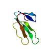
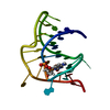
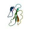
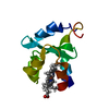
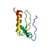
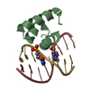
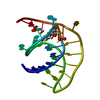
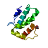
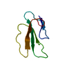
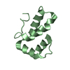
| Title | Structural Basis of the Membrane-induced Cardiotoxin A3 Oligomerization | ||||||
 Components Components | CARDIOTOXIN-3 | ||||||
 Keywords Keywords | CARDIOTOXIN / SODIUM DODECYL SULFATE / VENOM / CYTOTOXIN | ||||||
| Function / homology |  Function and homology information Function and homology informationcytolysis / other organism cell membrane / toxin activity / killing of cells of another organism / extracellular region / membrane Similarity search - Function | ||||||
| Biological species |  NAJA ATRA (Chinese cobra) NAJA ATRA (Chinese cobra) | ||||||
| Method |  X-RAY DIFFRACTION / MOLECULAR REPLACEMENT / Resolution: 1.9 Å X-RAY DIFFRACTION / MOLECULAR REPLACEMENT / Resolution: 1.9 Å | ||||||
 Authors Authors | Forouhar, F. / Huang, W.-N. / Liu, J.-H. / Chien, K.-Y. / Wu, W.-G. / Hsiao, C.-D. | ||||||
 Citation Citation | Journal: J.Biol.Chem. / Year: 2003 Title: Structural Basis of Membrane-Induced Cardiotoxin A3 Oligomerization Authors: Forouhar, F. / Huang, W.-N. / Liu, J.-H. / Chien, K.-Y. / Wu, W.-G. / Hsiao, C.-D. | ||||||
| History |
|
- Structure visualization
| Structure viewer | Molecule: MolmilJmol/JSmol |
|---|
- Downloads & links
-Download
| PDBx/mmCIF format | 1h0j.cif.gz | 55.3 KB | Display | PDBx/mmCIF format |
|---|---|---|---|---|
| PDB format | pdb1h0j.ent.gz | 42.8 KB | Display | PDB format |
| PDBx/mmJSON format | 1h0j.json.gz | Tree view | PDBx/mmJSON format | |
| Others |  Other downloads Other downloads |
-Validation report
| Arichive directory | https://data.pdbj.org/pub/pdb/validation_reports/h0/1h0jftp://data.pdbj.org/pub/pdb/validation_reports/h0/1h0j | HTTPS FTP |
|---|
-Related structure data
| Related structure data |  1tgxS S: Starting model for refinement |
|---|---|
| Similar structure data |


-Links
 PDBj
PDBj

- Assembly
| Deposited unit | 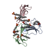
| ||||||||
|---|---|---|---|---|---|---|---|---|---|
| 1 | 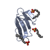
| ||||||||
| 2 | 
| ||||||||
| 3 | 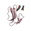
| ||||||||
| Unit cell |
|
-Components
| #1: Protein | Mass: 6758.330 Da / Num. of mol.: 3 / Source method: isolated from a natural source / Details: SODIUM DODECYL SULFATE / Source: (natural) NAJA ATRA (Chinese cobra) / Organ: VENOM GLAND / References: UniProt: P01444, UniProt: P60301*PLUS#2: Chemical | ChemComp-SDS /   Mass: 266.397 Da / Num. of mol.: 10 / Source method: obtained synthetically / Formula: C12H26O4S / Comment: detergent*YM Mass: 266.397 Da / Num. of mol.: 10 / Source method: obtained synthetically / Formula: C12H26O4S / Comment: detergent*YM#3: Water | ChemComp-HOH / |  Mass: 18.015 Da / Num. of mol.: 257 / Source method: isolated from a natural source / Formula: H2O Mass: 18.015 Da / Num. of mol.: 257 / Source method: isolated from a natural source / Formula: H2OCompound details | BELONGS TO THE SNAKE TOXIN FAMILY. | Has protein modification | Y | |
|---|
-Experimental details
-Experiment
| Experiment | Method: X-RAY DIFFRACTION / Number of used crystals: 1 |
|---|
- Sample preparation
| Crystal | Density Matthews: 2.36 Å3/Da / Density % sol: 48 % | ||||||||||||||||||||||||||||||||||||
|---|---|---|---|---|---|---|---|---|---|---|---|---|---|---|---|---|---|---|---|---|---|---|---|---|---|---|---|---|---|---|---|---|---|---|---|---|---|
| Crystal grow | pH: 4.6 Details: 10 MG/ML PROTEIN MIXED WITH 0.1M SODIUM ACETATE (PH 4.6), 20% PEG 400, 3% GLYCEROL, AND 24 MM SDS. | ||||||||||||||||||||||||||||||||||||
| Crystal grow | *PLUS pH: 4.6 / Method: vapor diffusion, hanging drop | ||||||||||||||||||||||||||||||||||||
| Components of the solutions | *PLUS
|
-Data collection
| Diffraction | Mean temperature: 110 K |
|---|---|
| Diffraction source | Source: ROTATING ANODE / Type: RIGAKU RU300 / Wavelength: 1.5418 |
| Detector | Type: RIGAKU IMAGE PLATE / Detector: IMAGE PLATE / Date: Sep 15, 2001 / Details: MIRRORS |
| Radiation | Protocol: SINGLE WAVELENGTH / Monochromatic (M) / Laue (L): M / Scattering type: x-ray |
| Radiation wavelength | Wavelength: 1.5418 Å / Relative weight: 1 |
| Reflection | Resolution: 1.9→30 Å / Num. obs: 21036 / % possible obs: 94.4 % / Observed criterion σ(I): 2 / Redundancy: 4.47 % / Biso Wilson estimate: 18.1 Å2 / Rmerge(I) obs: 0.046 / Net I/σ(I): 28.5 |
| Reflection shell | Resolution: 1.9→1.97 Å / Rmerge(I) obs: 0.266 / Mean I/σ(I) obs: 3.91 / % possible all: 96.3 |
| Reflection | *PLUS Highest resolution: 1.9 Å / Lowest resolution: 30 Å / Num. measured all: 93866 / Rmerge(I) obs: 0.046 |
| Reflection shell | *PLUS % possible obs: 96.3 % / Rmerge(I) obs: 0.266 |
- Processing
| Software |
| ||||||||||||||||||||||||||||||||||||||||||||||||||||||||||||
|---|---|---|---|---|---|---|---|---|---|---|---|---|---|---|---|---|---|---|---|---|---|---|---|---|---|---|---|---|---|---|---|---|---|---|---|---|---|---|---|---|---|---|---|---|---|---|---|---|---|---|---|---|---|---|---|---|---|---|---|---|---|
| Refinement | Method to determine structure: MOLECULAR REPLACEMENT Starting model: PDB ENTRY 1TGX Resolution: 1.9→30 Å / Rfactor Rfree error: 0.007 / Data cutoff high absF: 595624.89 / Isotropic thermal model: RESTRAINED / Cross valid method: THROUGHOUT / σ(F): 2
| ||||||||||||||||||||||||||||||||||||||||||||||||||||||||||||
| Solvent computation | Solvent model: FLAT MODEL / Bsol: 96.6429 Å2 / ksol: 0.389881 e/Å3 | ||||||||||||||||||||||||||||||||||||||||||||||||||||||||||||
| Displacement parameters | Biso mean: 33.8 Å2
| ||||||||||||||||||||||||||||||||||||||||||||||||||||||||||||
| Refine analyze |
| ||||||||||||||||||||||||||||||||||||||||||||||||||||||||||||
| Refinement step | Cycle: LAST / Resolution: 1.9→30 Å
| ||||||||||||||||||||||||||||||||||||||||||||||||||||||||||||
| Refine LS restraints |
| ||||||||||||||||||||||||||||||||||||||||||||||||||||||||||||
| LS refinement shell | Resolution: 1.9→2.02 Å / Rfactor Rfree error: 0.017 / Total num. of bins used: 6
| ||||||||||||||||||||||||||||||||||||||||||||||||||||||||||||
| Xplor file |
| ||||||||||||||||||||||||||||||||||||||||||||||||||||||||||||
| Refinement | *PLUS Rfactor Rfree: 0.2775 / Rfactor Rwork: 0.2201 | ||||||||||||||||||||||||||||||||||||||||||||||||||||||||||||
| Solvent computation | *PLUS | ||||||||||||||||||||||||||||||||||||||||||||||||||||||||||||
| Displacement parameters | *PLUS | ||||||||||||||||||||||||||||||||||||||||||||||||||||||||||||
| Refine LS restraints | *PLUS
|
