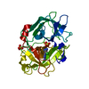
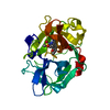
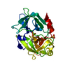
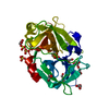
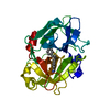
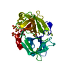
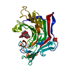
登録情報 データベース : PDB / ID : 1fv9タイトル Crystal structure of human microurokinase in complex with 2-amino-5-hydroxy-benzimidazole UROKINASE キーワード / 機能・相同性 分子機能 ドメイン・相同性 構成要素
/ / / / / / / / / / / / / / / / / / / / / / / / / / / / / / / / / / / / / / / / / / / / / / / / / / / / / / / / / / / / / / / / / / / / / / / / / / / / / / / / / / / 生物種 Homo sapiens (ヒト)手法 / 解像度 : 3 Å データ登録者 Nienaber, V. #1: ジャーナル : J.Biol.Chem. / 年 : 2000タイトル : Re-engineering of human urokinase provides a system for structure-based drug design at high resolution and reveals a novel structural subsite
履歴 登録 2000年9月19日 登録サイト / 処理サイト 改定 1.0 2000年10月18日 Provider / タイプ 改定 1.1 2008年4月27日 Group 改定 1.2 2011年7月13日 Group 改定 1.3 2017年10月4日 Group / カテゴリ 改定 1.4 2018年1月31日 Group / カテゴリ / Item 改定 1.5 2024年11月13日 Group Data collection / Database references ... Data collection / Database references / Derived calculations / Structure summary カテゴリ chem_comp_atom / chem_comp_bond ... chem_comp_atom / chem_comp_bond / database_2 / pdbx_entry_details / pdbx_modification_feature / struct_ref_seq_dif / struct_site Item _database_2.pdbx_DOI / _database_2.pdbx_database_accession ... _database_2.pdbx_DOI / _database_2.pdbx_database_accession / _struct_ref_seq_dif.details / _struct_site.pdbx_auth_asym_id / _struct_site.pdbx_auth_comp_id / _struct_site.pdbx_auth_seq_id
すべて表示 表示を減らす
 ムービー
ムービー コントローラー


 データを開く
 基本情報
基本情報 要素
要素 キーワード
キーワード 機能・相同性情報
 Homo sapiens (ヒト)
 X線回折 / 解像度: 3 Å
  データ登録者
 引用
引用 構造の表示
構造の表示 ダウンロードとリンク
 その他のダウンロード
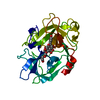


 PDBj
PDBj

 集合体
集合体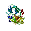

 Spodoptera frugiperda (ツマジロクサヨトウ)
 分子量: 96.063 Da / 分子数: 1 / 由来タイプ: 合成 / 式: SO4
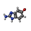
 分子量: 149.150 Da / 分子数: 1 / 由来タイプ: 合成 / 式: C7H7N3O
 試料調製
試料調製 解析
解析