 Movie
Movie Controller

[English] 日本語
 Yorodumi
- PDB-1e2i: The nucleoside binding site of Herpes simplex type 1 thymidine ki... -
+ Open data

- Basic information
| Entry | Database: PDB / ID: 1e2i | ||||||
|---|---|---|---|---|---|---|---|
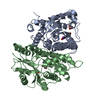
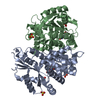
| Title | The nucleoside binding site of Herpes simplex type 1 thymidine kinase analyzed by X-ray crystallography | ||||||
 Components Components | THYMIDINE KINASE | ||||||
 Keywords Keywords | TRANSFERASE / ADENINE ANALOG / ENZYME-PRODRUG GENE THERAPY / NUCLEOSIDE- BINDING / THYMIDINE KINASE | ||||||
| Function / homology |  Function and homology information Function and homology informationTMP biosynthetic process / thymidine kinase / thymidine kinase activity / DNA biosynthetic process / ATP binding Similarity search - Function | ||||||
| Biological species |  HERPES SIMPLEX VIRUS HERPES SIMPLEX VIRUS | ||||||
| Method |  X-RAY DIFFRACTION / SYNCHROTRON / MOLECULAR REPLACEMENT / Resolution: 1.9 Å X-RAY DIFFRACTION / SYNCHROTRON / MOLECULAR REPLACEMENT / Resolution: 1.9 Å | ||||||
 Authors Authors | Vogt, J. / Scapozza, L. / Schulz, G.E. | ||||||
 Citation Citation | Journal: Proteins / Year: 2000 Title: Nucleoside Binding Site of Herpes Simplex Type 1 Thymidine Kinase Analyzed by X-Ray Crystallography Authors: Vogt, J. / Perozzo, R. / Pautsch, A. / Prota, A. / Schelling, P. / Pilger, B. / Folkers, G. / Scapozza, L. / Schulz, G.E. #1: Journal: FEBS Lett. / Year: 1995 Title: The Three-Dimensional Structure of Thymidine Kinase from Herpes Simplex Virus Type 1 Authors: Wild, K. / Bohner, T. / Aubry, A. / Folkers, G. / Schulz, G.E. #2: Journal: Nat.Struct.Biol. / Year: 1995 Title: Crystal Structures of the Thymidine Kinase from Herpes Simplex Virus Type-1 in Complex with Deoxythymidine and Ganciclovir Authors: Brown, D.G. / Visse, R. / Sandhu, G. / Davies, A. / Rizkallah, P.J. / Melitz, C. / Summers, W.C. / Sanderson, M.R. | ||||||
| History |
| ||||||
| Remark 700 | SHEET THE SHEET STRUCTURE OF THIS MOLECULE IS BIFURCATED. IN ORDER TO REPRESENT THIS FEATURE IN ... SHEET THE SHEET STRUCTURE OF THIS MOLECULE IS BIFURCATED. IN ORDER TO REPRESENT THIS FEATURE IN THE SHEET RECORDS BELOW, TWO SHEETS ARE DEFINED. |
- Structure visualization
| Structure viewer | Molecule: MolmilJmol/JSmol |
|---|
- Downloads & links
-Download
| PDBx/mmCIF format | 1e2i.cif.gz | 136.1 KB | Display | PDBx/mmCIF format |
|---|---|---|---|---|
| PDB format | pdb1e2i.ent.gz | 106.6 KB | Display | PDB format |
| PDBx/mmJSON format | 1e2i.json.gz | Tree view | PDBx/mmJSON format | |
| Others |  Other downloads Other downloads |
-Validation report
| Arichive directory | https://data.pdbj.org/pub/pdb/validation_reports/e2/1e2iftp://data.pdbj.org/pub/pdb/validation_reports/e2/1e2i | HTTPS FTP |
|---|
-Related structure data
| Related structure data | 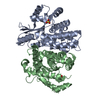 1e2hC  1e2jC 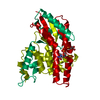 1vtkS S: Starting model for refinement C: citing same article ( |
|---|---|
| Similar structure data |


-Links
 PDBj
PDBj- Assembly
| Deposited unit | 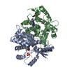
| ||||||||
|---|---|---|---|---|---|---|---|---|---|
| 1 |
| ||||||||
| Unit cell |
| ||||||||
| Components on special symmetry positions |
| ||||||||
| Noncrystallographic symmetry (NCS) | NCS oper: (Code: given Matrix: (-0.92993, -0.31266, -0.19356), Vector: Details | THE STRONG CRYSTAL PACKING GENERATED USING X , 1-Y, -Z GIVESCHAIN A TO CHAIN A (SYMMETRY RELATED) CONTACTS. | |
-Components
| #1: Protein | Mass: 35779.086 Da / Num. of mol.: 2 Source method: isolated from a genetically manipulated source Source: (gene. exp.) HERPES SIMPLEX VIRUS (TYPE 1 / STRAIN 17)Strain: 17 / Production host:  References: UniProt: P03176, UniProt: P0DTH5*PLUS, thymidine kinase #2: Chemical |   Mass: 96.063 Da / Num. of mol.: 2 / Source method: obtained synthetically / Formula: SO4 Mass: 96.063 Da / Num. of mol.: 2 / Source method: obtained synthetically / Formula: SO4#3: Chemical |   Mass: 193.206 Da / Num. of mol.: 2 / Source method: obtained synthetically / Formula: C8H11N5O Mass: 193.206 Da / Num. of mol.: 2 / Source method: obtained synthetically / Formula: C8H11N5O#4: Chemical |   Mass: 193.206 Da / Num. of mol.: 2 / Source method: obtained synthetically / Formula: C8H11N5O Mass: 193.206 Da / Num. of mol.: 2 / Source method: obtained synthetically / Formula: C8H11N5O#5: Water | ChemComp-HOH / |  Mass: 18.015 Da / Num. of mol.: 221 / Source method: isolated from a natural source / Formula: H2O Mass: 18.015 Da / Num. of mol.: 221 / Source method: isolated from a natural source / Formula: H2O |
|---|
-Experimental details
-Experiment
| Experiment | Method: X-RAY DIFFRACTION / Number of used crystals: 1 |
|---|
- Sample preparation
| Crystal | Density Matthews: 2.49 Å3/Da / Density % sol: 50.64 % | ||||||||||||||||||||||||||||||
|---|---|---|---|---|---|---|---|---|---|---|---|---|---|---|---|---|---|---|---|---|---|---|---|---|---|---|---|---|---|---|---|
| Crystal grow | pH: 7.5 / Details: LITHIUM SULFATE, HEPES, DTT, pH 7.50 | ||||||||||||||||||||||||||||||
| Crystal grow | *PLUS Temperature: 23 ℃ / Method: vapor diffusion, sitting drop / PH range low: 8 / PH range high: 7.5 | ||||||||||||||||||||||||||||||
| Components of the solutions | *PLUS
|
-Data collection
| Diffraction | Mean temperature: 100 K |
|---|---|
| Diffraction source | Source: SYNCHROTRON / Site: EMBL/DESY, HAMBURG  / Beamline: BW7B / Wavelength: 0.8468 / Beamline: BW7B / Wavelength: 0.8468 |
| Detector | Type: MARRESEARCH / Detector: IMAGE PLATE / Date: Mar 12, 1999 |
| Radiation | Protocol: SINGLE WAVELENGTH / Monochromatic (M) / Laue (L): M / Scattering type: x-ray |
| Radiation wavelength | Wavelength: 0.8468 Å / Relative weight: 1 |
| Reflection | Resolution: 1.9→20 Å / Num. obs: 54250 / % possible obs: 96 % / Redundancy: 3.3 % / Biso Wilson estimate: 29.8 Å2 / Rmerge(I) obs: 0.05 / Rsym value: 0.05 / Net I/σ(I): 12.5 |
| Reflection shell | Resolution: 1.9→2 Å / Redundancy: 2.8 % / Rmerge(I) obs: 0.425 / Mean I/σ(I) obs: 2.7 / Rsym value: 0.425 / % possible all: 96 |
| Reflection | *PLUS Rmerge(I) obs: 0.05 |
- Processing
| Software |
| |||||||||||||||||||||||||||||||||||||||||||||||||||||||||||||||
|---|---|---|---|---|---|---|---|---|---|---|---|---|---|---|---|---|---|---|---|---|---|---|---|---|---|---|---|---|---|---|---|---|---|---|---|---|---|---|---|---|---|---|---|---|---|---|---|---|---|---|---|---|---|---|---|---|---|---|---|---|---|---|---|---|
| Refinement | Method to determine structure: MOLECULAR REPLACEMENT Starting model: 1VTK Resolution: 1.9→20 Å / SU B: 2.4 / SU ML: 0.07 / Cross valid method: THROUGHOUT / σ(F): 0 / ESU R: 0.16 / ESU R Free: 0.16
| |||||||||||||||||||||||||||||||||||||||||||||||||||||||||||||||
| Displacement parameters | Biso mean: 42 Å2 | |||||||||||||||||||||||||||||||||||||||||||||||||||||||||||||||
| Refinement step | Cycle: LAST / Resolution: 1.9→20 Å
| |||||||||||||||||||||||||||||||||||||||||||||||||||||||||||||||
| Refine LS restraints |
| |||||||||||||||||||||||||||||||||||||||||||||||||||||||||||||||
| Software | *PLUS Name: REFMAC / Classification: refinement | |||||||||||||||||||||||||||||||||||||||||||||||||||||||||||||||
| Refinement | *PLUS Rfactor obs: 0.222 | |||||||||||||||||||||||||||||||||||||||||||||||||||||||||||||||
| Solvent computation | *PLUS | |||||||||||||||||||||||||||||||||||||||||||||||||||||||||||||||
| Displacement parameters | *PLUS |
