 Movie
Movie Controller


+ Open data

- Basic information
| Entry | Database: PDB / ID: 1duh | ||||||
|---|---|---|---|---|---|---|---|
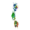
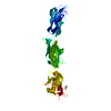
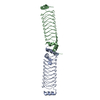
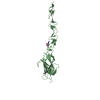
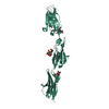
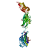
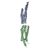
| Title | CRYSTAL STRUCTURE OF THE CONSERVED DOMAIN IV OF E. COLI 4.5S RNA | ||||||
 Components Components | 4.5S RNA DOMAIN IV | ||||||
 Keywords Keywords | RNA / 4.5S RNA / DOMAIN IV / HELIX 8 / SIGNAL RECOGNITION PARTICLE / SRP / FFH / SRP54 / ELONGATION FACTOR G / EF-G / 23S RNA / NON-CANONICAL BASE PAIRS / MISMATCH | ||||||
| Function / homology | : / : / RNA / RNA (> 10) Function and homology information Function and homology information | ||||||
| Method |  X-RAY DIFFRACTION / SYNCHROTRON / MAD / Resolution: 2.7 Å X-RAY DIFFRACTION / SYNCHROTRON / MAD / Resolution: 2.7 Å | ||||||
 Authors Authors | Jovine, L. / Hainzl, T. / Oubridge, C. / Scott, W.G. / Li, J. / Sixma, T.K. / Wonacott, A. / Skarzynski, T. / Nagai, K. | ||||||
 Citation Citation | Journal: Structure Fold.Des. / Year: 2000 Title: Crystal structure of the ffh and EF-G binding sites in the conserved domain IV of Escherichia coli 4.5S RNA. Authors: Jovine, L. / Hainzl, T. / Oubridge, C. / Scott, W.G. / Li, J. / Sixma, T.K. / Wonacott, A. / Skarzynski, T. / Nagai, K. #1: Journal: Acta Crystallogr.,Sect.D / Year: 2000Title: Crystallization and preliminary X-ray analysis of the conserved domain IV of E. coli 4.5S RNA Authors: Jovine, L. / Hainzl, T. / Oubridge, C. / Nagai, K. | ||||||
| History |
|
- Structure visualization
| Structure viewer | Molecule: MolmilJmol/JSmol |
|---|
- Downloads & links
-Download
| PDBx/mmCIF format | 1duh.cif.gz | 38.9 KB | Display | PDBx/mmCIF format |
|---|---|---|---|---|
| PDB format | pdb1duh.ent.gz | 25 KB | Display | PDB format |
| PDBx/mmJSON format | 1duh.json.gz | Tree view | PDBx/mmJSON format | |
| Others |  Other downloads Other downloads |
-Validation report
| Arichive directory | https://data.pdbj.org/pub/pdb/validation_reports/du/1duhftp://data.pdbj.org/pub/pdb/validation_reports/du/1duh | HTTPS FTP |
|---|
-Related structure data
| Similar structure data |
|---|

-Links
 PDBj
PDBj


- Assembly
| Deposited unit | 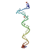
| ||||||||||
|---|---|---|---|---|---|---|---|---|---|---|---|
| 1 | 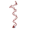
| ||||||||||
| Unit cell |
|
-Components
| #1: RNA chain | Mass: 14663.736 Da / Num. of mol.: 1 / Fragment: DOMAIN IV / Source method: obtained synthetically Details: RNA SEQUENCE TAKEN FROM ESCHERICHIA COLI 4.5S RNA. THE RNA WAS PRODUCED BY T7 RNA POLYMERASE IN VITRO TRANSCRIPTION USING RIBOZYME TECHNOLOGY References: EMBL: X01074 | ||
|---|---|---|---|
| #2: Chemical | ChemComp-LU /   Mass: 174.967 Da / Num. of mol.: 1 / Source method: obtained synthetically / Formula: Lu Mass: 174.967 Da / Num. of mol.: 1 / Source method: obtained synthetically / Formula: Lu | ||
| #3: Chemical | ChemComp-MG /   Mass: 24.305 Da / Num. of mol.: 1 / Source method: obtained synthetically / Formula: Mg Mass: 24.305 Da / Num. of mol.: 1 / Source method: obtained synthetically / Formula: Mg | ||
| #4: Chemical |   Mass: 96.063 Da / Num. of mol.: 2 / Source method: obtained synthetically / Formula: SO4 Mass: 96.063 Da / Num. of mol.: 2 / Source method: obtained synthetically / Formula: SO4#5: Water | ChemComp-HOH / |  Mass: 18.015 Da / Num. of mol.: 6 / Source method: isolated from a natural source / Formula: H2O Mass: 18.015 Da / Num. of mol.: 6 / Source method: isolated from a natural source / Formula: H2O |
-Experimental details
-Experiment
| Experiment | Method: X-RAY DIFFRACTION / Number of used crystals: 1 |
|---|
- Sample preparation
| Crystal | Density Matthews: 4.15 Å3/Da / Density % sol: 76.9 % | ||||||||||||||||||||||||||||||||||||||||||||||||||||||
|---|---|---|---|---|---|---|---|---|---|---|---|---|---|---|---|---|---|---|---|---|---|---|---|---|---|---|---|---|---|---|---|---|---|---|---|---|---|---|---|---|---|---|---|---|---|---|---|---|---|---|---|---|---|---|---|
| Crystal grow | Temperature: 303.15 K / Method: vapor diffusion, sitting drop / pH: 6 Details: CRYSTALLIZED FROM 1.60-1.90 M (NH4)2SO4, 0.09 M MAGNESIUM ACETATE, 0.05 M SODIUM CACODYLATE PH 6.0, AT 303 K. CRYSTALS WERE STABILISED AT 292 K IN A SOLUTION OF 2.20 M (NH4)2SO4, 0.01 M ...Details: CRYSTALLIZED FROM 1.60-1.90 M (NH4)2SO4, 0.09 M MAGNESIUM ACETATE, 0.05 M SODIUM CACODYLATE PH 6.0, AT 303 K. CRYSTALS WERE STABILISED AT 292 K IN A SOLUTION OF 2.20 M (NH4)2SO4, 0.01 M MGCL2, 0.05 M BIS-TRIS-HCL PH 6.0. SOAK CONDITIONS: CRYSTALS WERE SOAKED IN STABILISATION SOLUTION CONTAINING 0.002 M LUTETIUM CHLORIDE HEXAHYDRATE. CRYOPROTECTION CONDITIONS: AFTER ADDITION OF 20% GLYCEROL (W/V) TO THE SOAK SOLUTION, CRYSTALS WERE FLASH-FROZEN IN LIQUID NITROGEN., VAPOR DIFFUSION, SITTING DROP, temperature 303.15K | ||||||||||||||||||||||||||||||||||||||||||||||||||||||
| Components of the solutions |
| ||||||||||||||||||||||||||||||||||||||||||||||||||||||
| Crystal grow | *PLUS | ||||||||||||||||||||||||||||||||||||||||||||||||||||||
| Components of the solutions | *PLUS
|
-Data collection
| Diffraction | Mean temperature: 100 K | ||||||||||||||||||
|---|---|---|---|---|---|---|---|---|---|---|---|---|---|---|---|---|---|---|---|
| Diffraction source | Source: SYNCHROTRON / Site: ELETTRA  / Beamline: 5.2R / Wavelength: 1.0302, 1.3366, 1.3369, 0.9968, 1.3359 / Beamline: 5.2R / Wavelength: 1.0302, 1.3366, 1.3369, 0.9968, 1.3359 | ||||||||||||||||||
| Detector | Type: MARRESEARCH / Detector: IMAGE PLATE / Date: Aug 1, 1999 | ||||||||||||||||||
| Radiation | Protocol: MAD / Monochromatic (M) / Laue (L): M / Scattering type: x-ray | ||||||||||||||||||
| Radiation wavelength |
| ||||||||||||||||||
| Reflection | Resolution: 2.7→22.5 Å / Num. all: 12371 / Num. obs: 12371 / % possible obs: 98.5 % / Observed criterion σ(F): 0 / Observed criterion σ(I): 0 / Redundancy: 4.2 % / Biso Wilson estimate: 100.3 Å2 / Rmerge(I) obs: 0.076 / Net I/σ(I): 14.5 | ||||||||||||||||||
| Reflection shell | Resolution: 2.7→2.8 Å / Redundancy: 4.1 % / Rmerge(I) obs: 0.593 / Mean I/σ(I) obs: 2.1 / % possible all: 100 | ||||||||||||||||||
| Reflection | *PLUS Num. measured all: 154595 | ||||||||||||||||||
| Reflection shell | *PLUS % possible obs: 100 % |
- Processing
| Software |
| ||||||||||||||||||||||||||||||||||||||||||||||||||||||||||||||||||||||||
|---|---|---|---|---|---|---|---|---|---|---|---|---|---|---|---|---|---|---|---|---|---|---|---|---|---|---|---|---|---|---|---|---|---|---|---|---|---|---|---|---|---|---|---|---|---|---|---|---|---|---|---|---|---|---|---|---|---|---|---|---|---|---|---|---|---|---|---|---|---|---|---|---|---|
| Refinement | Method to determine structure: MAD / Resolution: 2.7→22.5 Å / Rfactor Rfree error: 0.008 / Data cutoff high absF: 1101782.89 / Data cutoff low absF: 0 / Isotropic thermal model: RESTRAINED / Cross valid method: THROUGHOUT / σ(F): 0 / σ(I): 0 / Stereochemistry target values: PARKINSON ET AL. Details: A LOW RESOLUTION LIMIT OF 8.0 A WAS USED FOR INITIAL B FACTOR AND BULK SOLVENT CORRECTIONS. NUCLEOTIDE A39 WAS REFINED WITH OCCUPANCY OF 0.5 TO ACCOUNT FOR ITS ALTERNATIVELY ORDERED AND ...Details: A LOW RESOLUTION LIMIT OF 8.0 A WAS USED FOR INITIAL B FACTOR AND BULK SOLVENT CORRECTIONS. NUCLEOTIDE A39 WAS REFINED WITH OCCUPANCY OF 0.5 TO ACCOUNT FOR ITS ALTERNATIVELY ORDERED AND DISORDERED CONFORMATION IN ADJACENT MOLECULES WITHIN THE CRYSTAL. THE APPARENT DISCREPANCY BETWEEN DATA COMPLETENESS IN SCALING AND REFINEMENT IS DUE TO THE VERY HIGH ANISOTROPY OF THE CRYSTAL DIFFRACTION, SO THAT, ALTHOUGH DATA COVERAGE WAS COMPLETE UP TO 2.70 A RESOLUTION, A SIGNIFICANT PROPORTION OF REFLECTIONS AT HIGH RESOLUTION WERE EXTINCT. THIS RESULTS IN THE HIGH R SYM IN THE OUTER SHELL, AND THE LOWER EFFECTIVE DATA COMPLETENESS DURING REFINEMENT (EXTINCT REFLECTIONS WERE OMITTED BY CNS).
| ||||||||||||||||||||||||||||||||||||||||||||||||||||||||||||||||||||||||
| Solvent computation | Solvent model: FLAT MODEL / Bsol: 36.81 Å2 / ksol: 0.266 e/Å3 | ||||||||||||||||||||||||||||||||||||||||||||||||||||||||||||||||||||||||
| Displacement parameters | Biso mean: 80.6 Å2
| ||||||||||||||||||||||||||||||||||||||||||||||||||||||||||||||||||||||||
| Refine analyze |
| ||||||||||||||||||||||||||||||||||||||||||||||||||||||||||||||||||||||||
| Refinement step | Cycle: LAST / Resolution: 2.7→22.5 Å
| ||||||||||||||||||||||||||||||||||||||||||||||||||||||||||||||||||||||||
| Refine LS restraints |
| ||||||||||||||||||||||||||||||||||||||||||||||||||||||||||||||||||||||||
| LS refinement shell | Refine-ID: X-RAY DIFFRACTION / Total num. of bins used: 8
| ||||||||||||||||||||||||||||||||||||||||||||||||||||||||||||||||||||||||
| Xplor file |
|
