 Movie
Movie Controller

[English] 日本語
 Yorodumi
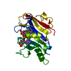
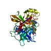
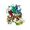
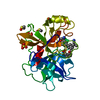
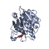
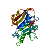
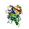
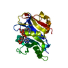
- PDB-1daj: COMPARISON OF TERNARY COMPLEXES OF PNEUMOCYSTIS CARINII AND WILD ... -
+ Open data

- Basic information
| Entry | Database: PDB / ID: 1daj | ||||||
|---|---|---|---|---|---|---|---|
| Title | COMPARISON OF TERNARY COMPLEXES OF PNEUMOCYSTIS CARINII AND WILD TYPE HUMAN DIHYDROFOLATE REDUCTASE WITH COENZYME NADPH AND A NOVEL CLASSICAL ANTITUMOR FURO[2,3D]PYRIMIDINE ANTIFOLATE | ||||||
 Components Components | DIHYDROFOLATE REDUCTASE | ||||||
 Keywords Keywords | OXIDOREDUCTASE | ||||||
| Function / homology |  Function and homology information Function and homology informationdihydrofolate metabolic process / glycine biosynthetic process / dihydrofolate reductase / dihydrofolate reductase activity / folic acid metabolic process / tetrahydrofolate biosynthetic process / one-carbon metabolic process / NADP binding / mitochondrion / cytosol Similarity search - Function | ||||||
| Biological species |  Pneumocystis carinii (fungus) Pneumocystis carinii (fungus) | ||||||
| Method |  X-RAY DIFFRACTION / Resolution: 2.3 Å X-RAY DIFFRACTION / Resolution: 2.3 Å | ||||||
 Authors Authors | Cody, V. / Galitsky, N. / Luft, J.R. / Pangborn, W. / Gangjee, A. / Devraj, R. / Queener, S.F. / Blakley, R.L. | ||||||
 Citation Citation | Journal: Acta Crystallogr.,Sect.D / Year: 1997 Title: Comparison of ternary complexes of Pneumocystis carinii and wild-type human dihydrofolate reductase with coenzyme NADPH and a novel classical antitumor furo[2,3-d]pyrimidine antifolate. Authors: Cody, V. / Galitsky, N. / Luft, J.R. / Pangborn, W. / Gangjee, A. / Devraj, R. / Queener, S.F. / Blakley, R.L. #1: Journal: Structure / Year: 1994Title: The Structure of Pneumocystis Carinii Dihydrofolate Reductase to 1.9 A Resolution Authors: Champness, J.N. / Achari, A. / Ballantine, S.P. / Bryant, P.K. / Delves, C.J. / Stammers, D.K. | ||||||
| History |
|
- Structure visualization
| Structure viewer | Molecule: MolmilJmol/JSmol |
|---|
- Downloads & links
-Download
| PDBx/mmCIF format | 1daj.cif.gz | 58 KB | Display | PDBx/mmCIF format |
|---|---|---|---|---|
| PDB format | pdb1daj.ent.gz | 41.6 KB | Display | PDB format |
| PDBx/mmJSON format | 1daj.json.gz | Tree view | PDBx/mmJSON format | |
| Others |  Other downloads Other downloads |
-Validation report
| Summary document | 1daj_validation.pdf.gz | 530.8 KB | Display | wwPDB validaton report |
|---|---|---|---|---|
| Full document | 1daj_full_validation.pdf.gz | 550.9 KB | Display | |
| Data in XML | 1daj_validation.xml.gz | 9.7 KB | Display | |
| Data in CIF | 1daj_validation.cif.gz | 12.9 KB | Display | |
| Arichive directory | https://data.pdbj.org/pub/pdb/validation_reports/da/1dajftp://data.pdbj.org/pub/pdb/validation_reports/da/1daj | HTTPS FTP |
-Related structure data
| Similar structure data |
|---|


-Links
 PDBj
PDBj

- Assembly
| Deposited unit | 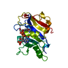
| ||||||||
|---|---|---|---|---|---|---|---|---|---|
| 1 |
| ||||||||
| Unit cell |
|
-Components
| #1: Protein | Mass: 23918.537 Da / Num. of mol.: 1 Source method: isolated from a genetically manipulated source Source: (gene. exp.) Pneumocystis carinii (fungus) / Gene: C-DNA PNEUMOCYSTIS CARINII DHF / Plasmid: PT7-7 / Gene (production host): C-DNA PNEUMOCYSTIS CARINII DHFR / Production host:  |
|---|---|
| #2: Chemical | ChemComp-NDP /   Mass: 745.421 Da / Num. of mol.: 1 / Source method: obtained synthetically / Formula: C21H30N7O17P3 Mass: 745.421 Da / Num. of mol.: 1 / Source method: obtained synthetically / Formula: C21H30N7O17P3 |
| #3: Chemical | ChemComp-MOT / 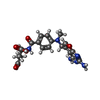  Mass: 442.425 Da / Num. of mol.: 1 / Source method: obtained synthetically / Formula: C20H22N6O6 Mass: 442.425 Da / Num. of mol.: 1 / Source method: obtained synthetically / Formula: C20H22N6O6 |
| #4: Water | ChemComp-HOH /  Mass: 18.015 Da / Num. of mol.: 50 / Source method: isolated from a natural source / Formula: H2O Mass: 18.015 Da / Num. of mol.: 50 / Source method: isolated from a natural source / Formula: H2O |
-Experimental details
-Experiment
| Experiment | Method: X-RAY DIFFRACTION |
|---|
- Sample preparation
| Crystal | Density Matthews: 2.04 Å3/Da / Density % sol: 39.6 % | ||||||||||||||||||||||||||||||||||||||||
|---|---|---|---|---|---|---|---|---|---|---|---|---|---|---|---|---|---|---|---|---|---|---|---|---|---|---|---|---|---|---|---|---|---|---|---|---|---|---|---|---|---|
| Crystal grow | *PLUS pH: 7 / Method: vapor diffusion, hanging drop | ||||||||||||||||||||||||||||||||||||||||
| Components of the solutions | *PLUS
|
-Data collection
| Diffraction source | Wavelength: 1.5418 |
|---|---|
| Detector | Type: RIGAKU RAXIS / Detector: IMAGE PLATE / Date: Jul 14, 1994 |
| Radiation | Scattering type: x-ray |
| Radiation wavelength | Wavelength: 1.5418 Å / Relative weight: 1 |
| Reflection | Num. obs: 5958 / % possible obs: 65.5 % / Observed criterion σ(I): 2 / Redundancy: 3.09 % / Rmerge(I) obs: 0.07 |
- Processing
| Software |
| ||||||||||||||||||||||||||||||||||||||||||||||||||||||||||||||||||||||||||||||||||||
|---|---|---|---|---|---|---|---|---|---|---|---|---|---|---|---|---|---|---|---|---|---|---|---|---|---|---|---|---|---|---|---|---|---|---|---|---|---|---|---|---|---|---|---|---|---|---|---|---|---|---|---|---|---|---|---|---|---|---|---|---|---|---|---|---|---|---|---|---|---|---|---|---|---|---|---|---|---|---|---|---|---|---|---|---|---|
| Refinement | Resolution: 2.3→8 Å /
| ||||||||||||||||||||||||||||||||||||||||||||||||||||||||||||||||||||||||||||||||||||
| Displacement parameters | Biso mean: 26.72 Å2 | ||||||||||||||||||||||||||||||||||||||||||||||||||||||||||||||||||||||||||||||||||||
| Refinement step | Cycle: LAST / Resolution: 2.3→8 Å
| ||||||||||||||||||||||||||||||||||||||||||||||||||||||||||||||||||||||||||||||||||||
| Refine LS restraints |
| ||||||||||||||||||||||||||||||||||||||||||||||||||||||||||||||||||||||||||||||||||||
| Software | *PLUS Name: PROFFT / Classification: refinement | ||||||||||||||||||||||||||||||||||||||||||||||||||||||||||||||||||||||||||||||||||||
| Refinement | *PLUS Rfactor obs: 0.181 | ||||||||||||||||||||||||||||||||||||||||||||||||||||||||||||||||||||||||||||||||||||
| Solvent computation | *PLUS | ||||||||||||||||||||||||||||||||||||||||||||||||||||||||||||||||||||||||||||||||||||
| Displacement parameters | *PLUS |
