[English] 日本語
 Yorodumi
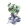
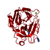
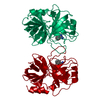
- PDB-1d7d: CYTOCHROME DOMAIN OF CELLOBIOSE DEHYDROGENASE, HP3 FRAGMENT, PH 7.5 -
+ Open data
- Basic information
| Entry | Database: PDB / ID: 1d7d | ||||||||||||
|---|---|---|---|---|---|---|---|---|---|---|---|---|---|
| Title | CYTOCHROME DOMAIN OF CELLOBIOSE DEHYDROGENASE, HP3 FRAGMENT, PH 7.5 | ||||||||||||
 Components Components | CELLOBIOSE DEHYDROGENASE | ||||||||||||
 Keywords Keywords | OXIDOREDUCTASE / B-TYPE CYTOCHROME / MET/HIS LIGATION / BETA SANDWICH / FE(II)-PROTOPORPHYRIN IX | ||||||||||||
| Function / homology |  Function and homology information Function and homology informationcellobiose dehydrogenase (acceptor) / cellobiose dehydrogenase (acceptor) activity / cellulose catabolic process / flavin adenine dinucleotide binding / extracellular region / metal ion binding Similarity search - Function | ||||||||||||
| Biological species |  Phanerochaete chrysosporium (fungus) Phanerochaete chrysosporium (fungus) | ||||||||||||
| Method |  X-RAY DIFFRACTION / X-RAY DIFFRACTION /  SYNCHROTRON / Resolution: 1.9 Å SYNCHROTRON / Resolution: 1.9 Å | ||||||||||||
 Authors Authors | Hallberg, B.M. / Bergfors, T. / Backbro, K. / Divne, C. | ||||||||||||
 Citation Citation |  Journal: Structure Fold.Des. / Year: 2000 Journal: Structure Fold.Des. / Year: 2000Title: A new scaffold for binding haem in the cytochrome domain of the extracellular flavocytochrome cellobiose dehydrogenase. Authors: Hallberg, B.M. / Bergfors, T. / Backbro, K. / Pettersson, G. / Henriksson, G. / Divne, C. | ||||||||||||
| History |
|
- Structure visualization
| Structure viewer | Molecule:  Molmil Molmil Jmol/JSmol Jmol/JSmol |
|---|
- Downloads & links
- Download
| PDBx/mmCIF format |  1d7d.cif.gz 1d7d.cif.gz | 96.8 KB | Display |  PDBx/mmCIF format PDBx/mmCIF format |
|---|---|---|---|---|
| PDB format |  pdb1d7d.ent.gz pdb1d7d.ent.gz | 72.9 KB | Display |  PDB format PDB format |
| PDBx/mmJSON format |  1d7d.json.gz 1d7d.json.gz | Tree view |  PDBx/mmJSON format PDBx/mmJSON format | |
| Others |  Other downloads Other downloads |
-Validation report
| Arichive directory |  https://data.pdbj.org/pub/pdb/validation_reports/d7/1d7d https://data.pdbj.org/pub/pdb/validation_reports/d7/1d7d ftp://data.pdbj.org/pub/pdb/validation_reports/d7/1d7d ftp://data.pdbj.org/pub/pdb/validation_reports/d7/1d7d | HTTPS FTP |
|---|
-Related structure data
- Links
Links
- Assembly
| Deposited unit | 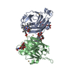
| ||||||||
|---|---|---|---|---|---|---|---|---|---|
| 1 |
| ||||||||
| Unit cell |
| ||||||||
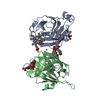
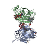
| Details | THE BIOLOGICAL ASSEMBLY IS A MONOMERIC FLAVOCYTOCHROME CONSISTING OF A B-TYPE CYTOCHROME DOMAIN LINKED TO A FLAVODEHYDROGENASE DOMAIN. |
- Components
-Protein , 1 types, 2 molecules AB
| #1: Protein | Mass: 20012.768 Da / Num. of mol.: 2 / Fragment: CYTOCHROME TYPE B DOMAIN / Source method: isolated from a natural source / Source: (natural)  Phanerochaete chrysosporium (fungus) / Tissue: SECRETED Phanerochaete chrysosporium (fungus) / Tissue: SECRETEDReferences: GenBank: 1314367, UniProt: Q01738*PLUS, EC: 1.1.3.25 |
|---|
-Sugars , 2 types, 2 molecules
| #2: Polysaccharide | beta-D-mannopyranose-(1-4)-2-acetamido-2-deoxy-beta-D-glucopyranose-(1-4)-2-acetamido-2-deoxy-beta- ...beta-D-mannopyranose-(1-4)-2-acetamido-2-deoxy-beta-D-glucopyranose-(1-4)-2-acetamido-2-deoxy-beta-D-glucopyranose Source method: isolated from a genetically manipulated source |
|---|---|
| #3: Polysaccharide | alpha-D-mannopyranose-(1-4)-2-acetamido-2-deoxy-beta-D-glucopyranose-(1-4)-2-acetamido-2-deoxy-beta- ...alpha-D-mannopyranose-(1-4)-2-acetamido-2-deoxy-beta-D-glucopyranose-(1-4)-2-acetamido-2-deoxy-beta-D-glucopyranose Source method: isolated from a genetically manipulated source |
-Non-polymers , 4 types, 278 molecules 

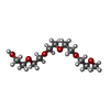


| #4: Chemical | ChemComp-CD / #5: Chemical | #6: Chemical | ChemComp-1PG / | #7: Water | ChemComp-HOH / | |
|---|
-Details
| Has protein modification | Y |
|---|
-Experimental details
-Experiment
| Experiment | Method:  X-RAY DIFFRACTION / Number of used crystals: 1 X-RAY DIFFRACTION / Number of used crystals: 1 |
|---|
- Sample preparation
| Crystal | Density Matthews: 3.68 Å3/Da / Density % sol: 66.58 % | ||||||||||||||||||||||||||||||||||||
|---|---|---|---|---|---|---|---|---|---|---|---|---|---|---|---|---|---|---|---|---|---|---|---|---|---|---|---|---|---|---|---|---|---|---|---|---|---|
| Crystal grow | Temperature: 298 K / Method: vapor diffusion, hanging drop / pH: 7.5 Details: PEG 4000, 2-methyl-2,4-pentanediol, hepes, cadmium chloride, pH 7.5, VAPOR DIFFUSION, HANGING DROP, temperature 298.0K | ||||||||||||||||||||||||||||||||||||
| Crystal grow | *PLUS Details: drop consists of equal amounts of protein and reservoir solutions | ||||||||||||||||||||||||||||||||||||
| Components of the solutions | *PLUS
|
-Data collection
| Diffraction | Mean temperature: 100 K |
|---|---|
| Diffraction source | Source:  SYNCHROTRON / Site: SYNCHROTRON / Site:  MAX II MAX II  / Beamline: I711 / Wavelength: 1.282 / Beamline: I711 / Wavelength: 1.282 |
| Detector | Type: MARRESEARCH / Detector: IMAGE PLATE / Date: Nov 24, 1998 |
| Radiation | Protocol: SINGLE WAVELENGTH / Monochromatic (M) / Laue (L): M / Scattering type: x-ray |
| Radiation wavelength | Wavelength: 1.282 Å / Relative weight: 1 |
| Reflection | Resolution: 1.9→25 Å / Num. all: 45909 / Num. obs: 45909 / % possible obs: 99.4 % / Observed criterion σ(F): 0 / Observed criterion σ(I): 0 / Redundancy: 5.2 % / Biso Wilson estimate: 18 Å2 / Rmerge(I) obs: 0.084 / Net I/σ(I): 16.7 |
| Reflection shell | Resolution: 1.9→1.93 Å / Redundancy: 5.5 % / Rmerge(I) obs: 0.329 / Num. unique all: 2253 / % possible all: 100 |
| Reflection | *PLUS Num. measured all: 236651 |
| Reflection shell | *PLUS % possible obs: 100 % / Mean I/σ(I) obs: 5.6 |
- Processing
| Software |
| |||||||||||||||||||||||||
|---|---|---|---|---|---|---|---|---|---|---|---|---|---|---|---|---|---|---|---|---|---|---|---|---|---|---|
| Refinement | Resolution: 1.9→25 Å / σ(F): 0 / σ(I): 0 / Stereochemistry target values: ENGH & HUBER / Details: MAXIMUM LIKELIHOOD, BULK SOLVENT CORRECTION
| |||||||||||||||||||||||||
| Refinement step | Cycle: LAST / Resolution: 1.9→25 Å
| |||||||||||||||||||||||||
| Refine LS restraints |
| |||||||||||||||||||||||||
| Software | *PLUS Name: 'CNS, SHELXL-97' / Classification: refinement | |||||||||||||||||||||||||
| Refinement | *PLUS Num. reflection all: 43188 | |||||||||||||||||||||||||
| Solvent computation | *PLUS | |||||||||||||||||||||||||
| Displacement parameters | *PLUS | |||||||||||||||||||||||||
| Refine LS restraints | *PLUS
|
 Movie
Movie Controller







 PDBj
PDBj


