 Movie
Movie Controller

[English] 日本語
 Yorodumi
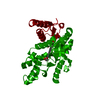
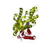
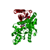
- PDB-1cet: CHLOROQUINE BINDS IN THE COFACTOR BINDING SITE OF PLASMODIUM FALC... -
+ Open data

- Basic information
| Entry | Database: PDB / ID: 1cet | ||||||
|---|---|---|---|---|---|---|---|
| Title | CHLOROQUINE BINDS IN THE COFACTOR BINDING SITE OF PLASMODIUM FALCIPARUM LACTATE DEHYDROGENASE. | ||||||
 Components Components | PROTEIN (L-LACTATE DEHYDROGENASE) | ||||||
 Keywords Keywords | OXIDOREDUCTASE / TRICARBOXYLIC ACID CYCLE / INHIBITOR | ||||||
| Function / homology |  Function and homology information Function and homology informationL-lactate dehydrogenase / L-lactate dehydrogenase (NAD+) activity / lactate metabolic process Similarity search - Function | ||||||
| Biological species |  | ||||||
| Method |  X-RAY DIFFRACTION / SYNCHROTRON / MOLECULAR REPLACEMENT / Resolution: 2.05 Å X-RAY DIFFRACTION / SYNCHROTRON / MOLECULAR REPLACEMENT / Resolution: 2.05 Å | ||||||
 Authors Authors | Read, J.A. / Wilkinson, K.W. / Tranter, R. / Sessions, R.B. / Brady, R.L. | ||||||
 Citation Citation | Journal: J.Biol.Chem. / Year: 1999 Title: Chloroquine binds in the cofactor binding site of Plasmodium falciparum lactate dehydrogenase. Authors: Read, J.A. / Wilkinson, K.W. / Tranter, R. / Sessions, R.B. / Brady, R.L. #1: Journal: Nat.Struct.Biol. / Year: 1996Title: The Structure of Lactate Dehydrogenase from Plasmodium Falciparum Reveals a New Target for Anti-Malarial Design Authors: Dunn, C.R. / Banfield, M.J. / Barker, J.J. / Higham, C.W. / Moreton, K.M. / Turgut-Balik, D. / Brady, R.L. / Holbrook, J.J. | ||||||
| History |
|
- Structure visualization
| Structure viewer | Molecule: MolmilJmol/JSmol |
|---|
- Downloads & links
-Download
| PDBx/mmCIF format | 1cet.cif.gz | 77.4 KB | Display | PDBx/mmCIF format |
|---|---|---|---|---|
| PDB format | pdb1cet.ent.gz | 55.8 KB | Display | PDB format |
| PDBx/mmJSON format | 1cet.json.gz | Tree view | PDBx/mmJSON format | |
| Others |  Other downloads Other downloads |
-Validation report
| Arichive directory | https://data.pdbj.org/pub/pdb/validation_reports/ce/1cetftp://data.pdbj.org/pub/pdb/validation_reports/ce/1cet | HTTPS FTP |
|---|
-Related structure data
| Related structure data | 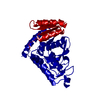 1ceqC 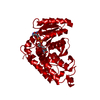 1ldgS S: Starting model for refinement C: citing same article ( |
|---|---|
| Similar structure data |


-Links
 PDBj
PDBj


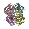
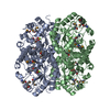
- Assembly
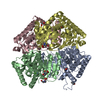
| Deposited unit | 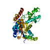
| ||||||||
|---|---|---|---|---|---|---|---|---|---|
| 1 | 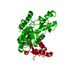
| ||||||||
| Unit cell |
|
-Components
| #1: Protein | Mass: 34162.785 Da / Num. of mol.: 1 Source method: isolated from a genetically manipulated source Source: (gene. exp.) Plasmid: PKK223-3 / Production host:  |
|---|---|
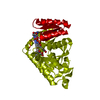
| #2: Chemical | ChemComp-CLQ / 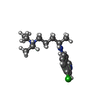  Mass: 319.872 Da / Num. of mol.: 1 / Source method: obtained synthetically / Formula: C18H26ClN3 / Comment: medication, antiparasitic*YM Mass: 319.872 Da / Num. of mol.: 1 / Source method: obtained synthetically / Formula: C18H26ClN3 / Comment: medication, antiparasitic*YM |
| #3: Water | ChemComp-HOH /  Mass: 18.015 Da / Num. of mol.: 252 / Source method: isolated from a natural source / Formula: H2O Mass: 18.015 Da / Num. of mol.: 252 / Source method: isolated from a natural source / Formula: H2O |
-Experimental details
-Experiment
| Experiment | Method: X-RAY DIFFRACTION / Number of used crystals: 1 |
|---|
- Sample preparation
| Crystal | Density Matthews: 2.3 Å3/Da / Density % sol: 40 % |
|---|---|
| Crystal grow | pH: 7.5 / Details: pH 7.5 |
-Data collection
| Diffraction | Mean temperature: 100 K |
|---|---|
| Diffraction source | Source: SYNCHROTRON / Site: SRS  / Beamline: PX7.2 / Wavelength: 1.488 / Beamline: PX7.2 / Wavelength: 1.488 |
| Detector | Type: MARRESEARCH / Detector: IMAGE PLATE |
| Radiation | Protocol: SINGLE WAVELENGTH / Monochromatic (M) / Laue (L): M / Scattering type: x-ray |
| Radiation wavelength | Wavelength: 1.488 Å / Relative weight: 1 |
| Reflection | Resolution: 2.05→20 Å / Num. obs: 20155 / % possible obs: 99.8 % / Observed criterion σ(I): 0 / Redundancy: 2.3 % / Rmerge(I) obs: 0.067 / Net I/σ(I): 16.3 |
- Processing
| Software |
| ||||||||||||||||||||||||||||||||||||||||||||||||||||||||||||
|---|---|---|---|---|---|---|---|---|---|---|---|---|---|---|---|---|---|---|---|---|---|---|---|---|---|---|---|---|---|---|---|---|---|---|---|---|---|---|---|---|---|---|---|---|---|---|---|---|---|---|---|---|---|---|---|---|---|---|---|---|---|
| Refinement | Method to determine structure: MOLECULAR REPLACEMENT Starting model: 1LDG Resolution: 2.05→20 Å / Data cutoff high absF: 20 / Data cutoff low absF: 2.05 / Cross valid method: THROUGHOUT / σ(F): 0
| ||||||||||||||||||||||||||||||||||||||||||||||||||||||||||||
| Displacement parameters | Biso mean: 9.1 Å2 | ||||||||||||||||||||||||||||||||||||||||||||||||||||||||||||
| Refinement step | Cycle: LAST / Resolution: 2.05→20 Å
| ||||||||||||||||||||||||||||||||||||||||||||||||||||||||||||
| Refine LS restraints |
| ||||||||||||||||||||||||||||||||||||||||||||||||||||||||||||
| Software | *PLUS Name: X-PLOR / Version: 3.851 / Classification: refinement | ||||||||||||||||||||||||||||||||||||||||||||||||||||||||||||
| Refinement | *PLUS Num. reflection all: 20115 / Num. reflection obs: 17783 | ||||||||||||||||||||||||||||||||||||||||||||||||||||||||||||
| Solvent computation | *PLUS | ||||||||||||||||||||||||||||||||||||||||||||||||||||||||||||
| Displacement parameters | *PLUS |
