 Movie
Movie Controller

[English] 日本語
 Yorodumi
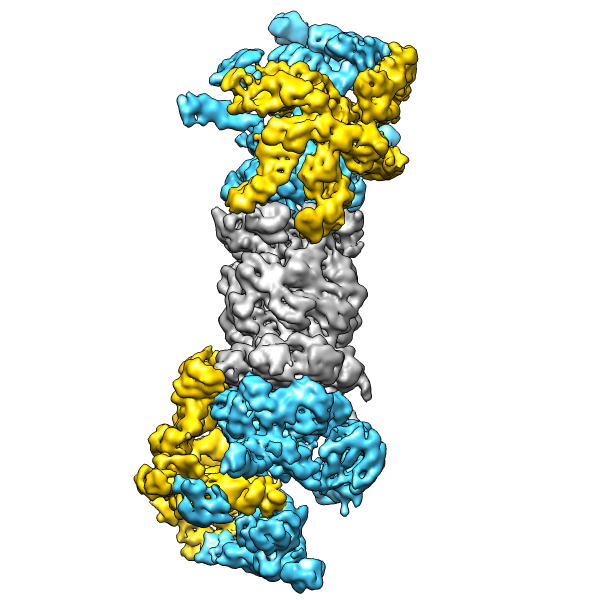
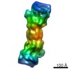
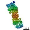
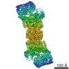
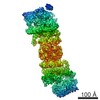
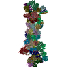
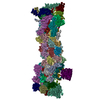
- EMDB-1992: The Saccharomyces cerevisiae 26S proteasome at subnanometer resolution -
+ Open data

- Basic information
| Entry | Database: EMDB / ID: EMD-1992 | |||||||||
|---|---|---|---|---|---|---|---|---|---|---|
| Title | The Saccharomyces cerevisiae 26S proteasome at subnanometer resolution | |||||||||
 Map data Map data | 26 proteasome | |||||||||
 Sample Sample |
| |||||||||
 Keywords Keywords | 26S / 19S / proteasome / regulatory particle / ubiquitin recognition / deubiquitination / AAA-ATPase | |||||||||
| Function / homology | proteasome regulatory particle / Proteasome, subunit alpha/beta Function and homology information Function and homology information | |||||||||
| Biological species |  | |||||||||
| Method | single particle reconstruction / cryo EM / negative staining / Resolution: 9.0 Å | |||||||||
 Authors Authors | Lander GC / Estrin E / Matyskiela M / Bashore C / Nogales E / Martin A | |||||||||
 Citation Citation | Journal: Nature / Year: 2012 Title: Complete subunit architecture of the proteasome regulatory particle. Authors: Gabriel C Lander / Eric Estrin / Mary E Matyskiela / Charlene Bashore / Eva Nogales / Andreas Martin /  Abstract: The proteasome is the major ATP-dependent protease in eukaryotic cells, but limited structural information restricts a mechanistic understanding of its activities. The proteasome regulatory particle, ...The proteasome is the major ATP-dependent protease in eukaryotic cells, but limited structural information restricts a mechanistic understanding of its activities. The proteasome regulatory particle, consisting of the lid and base subcomplexes, recognizes and processes polyubiquitinated substrates. Here we used electron microscopy and a new heterologous expression system for the lid to delineate the complete subunit architecture of the regulatory particle from yeast. Our studies reveal the spatial arrangement of ubiquitin receptors, deubiquitinating enzymes and the protein unfolding machinery at subnanometre resolution, outlining the substrate's path to degradation. Unexpectedly, the ATPase subunits within the base unfoldase are arranged in a spiral staircase, providing insight into potential mechanisms for substrate translocation through the central pore. Large conformational rearrangements of the lid upon holoenzyme formation suggest allosteric regulation of deubiquitination. We provide a structural basis for the ability of the proteasome to degrade a diverse set of substrates and thus regulate vital cellular processes. | |||||||||
| History |
|
- Structure visualization
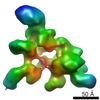
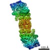
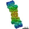
| Movie |
Movie viewer |
|---|---|
| Structure viewer | EM map: SurfViewMolmilJmol/JSmol |
| Supplemental images |
- Downloads & links
-EMDB archive
| Map data | emd_1992.map.gz | 2.8 MB | EMDB map data format | |
|---|---|---|---|---|
| Header (meta data) | emd-1992-v30.xmlemd-1992.xml | 10 KB 10 KB | Display Display | EMDB header |
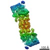
| Images |  emd-1992.png emd-1992.png | 199.4 KB | ||
| Archive directory |  http://ftp.pdbj.org/pub/emdb/structures/EMD-1992ftp://ftp.pdbj.org/pub/emdb/structures/EMD-1992 http://ftp.pdbj.org/pub/emdb/structures/EMD-1992ftp://ftp.pdbj.org/pub/emdb/structures/EMD-1992 | HTTPS FTP |
-Validation report
| Summary document | emd_1992_validation.pdf.gz | 208.3 KB | Display | EMDB validaton report |
|---|---|---|---|---|
| Full document | emd_1992_full_validation.pdf.gz | 207.4 KB | Display | |
| Data in XML | emd_1992_validation.xml.gz | 6.6 KB | Display | |
| Arichive directory | https://ftp.pdbj.org/pub/emdb/validation_reports/EMD-1992ftp://ftp.pdbj.org/pub/emdb/validation_reports/EMD-1992 | HTTPS FTP |
-Related structure data

-Links
| EMDB pages | EMDB (EBI/PDBe) / EMDataResource |
|---|
-Map
| File | Download / File: emd_1992.map.gz / Format: CCP4 / Size: 62.5 MB / Type: IMAGE STORED AS FLOATING POINT NUMBER (4 BYTES) | ||||||||||||||||||||||||||||||||||||||||||||||||||||||||||||||||||||
|---|---|---|---|---|---|---|---|---|---|---|---|---|---|---|---|---|---|---|---|---|---|---|---|---|---|---|---|---|---|---|---|---|---|---|---|---|---|---|---|---|---|---|---|---|---|---|---|---|---|---|---|---|---|---|---|---|---|---|---|---|---|---|---|---|---|---|---|---|---|
| Annotation | 26 proteasome | ||||||||||||||||||||||||||||||||||||||||||||||||||||||||||||||||||||
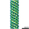
| Projections & slices | Image control
Images are generated by Spider. | ||||||||||||||||||||||||||||||||||||||||||||||||||||||||||||||||||||
| Voxel size | X=Y=Z: 2.17 Å | ||||||||||||||||||||||||||||||||||||||||||||||||||||||||||||||||||||
| Density |
| ||||||||||||||||||||||||||||||||||||||||||||||||||||||||||||||||||||
| Symmetry | Space group: 1 | ||||||||||||||||||||||||||||||||||||||||||||||||||||||||||||||||||||
| Details | EMDB XML:
CCP4 map header:
| ||||||||||||||||||||||||||||||||||||||||||||||||||||||||||||||||||||
 Z (Sec.)
 Y (Row.)
 X (Col.)


-Supplemental data
- Sample components
-Entire : 26S Proteasome
| Entire | Name: 26S Proteasome |
|---|---|
| Components |
|
-Supramolecule #1000: 26S Proteasome
| Supramolecule | Name: 26S Proteasome / type: sample / ID: 1000 / Details: monodisperse / Oligomeric state: holoenzyme / Number unique components: 1 |
|---|---|
| Molecular weight | Experimental: 1.5 MDa / Theoretical: 1.5 MDa / Method: Mass Spectrometry |
-Macromolecule #1: 26S Proteasome
| Macromolecule | Name: 26S Proteasome / type: protein_or_peptide / ID: 1 / Name.synonym: 26S Proteasome / Details: Endogenous proteasome was purified from yeast / Number of copies: 1 / Oligomeric state: monomer / Recombinant expression: No |
|---|---|
| Source (natural) | Organism: |
| Molecular weight | Experimental: 1.5 MDa / Theoretical: 1.5 MDa |
| Sequence | GO: proteasome regulatory particle / InterPro: Proteasome, subunit alpha/beta |
-Experimental details
-Structure determination
| Method | negative staining, cryo EM |
|---|---|
 Processing Processing | single particle reconstruction |
| Aggregation state | particle |
-Sample preparation
| Concentration | 1.7 mg/mL |
|---|---|
| Buffer | pH: 7.6 Details: 20mM HEPES, 50mM NaCl, 50mM KCl, 1mM ATP, 1mM DTT, 0.05% NP40 |
| Staining | Type: NEGATIVE Details: C-flat grids made hydrophilic with Solarus Plasma cleaner, 4 uL of sample applied, blotted in Vitrobot for 3 seconds with offset -1, plunged into liquid ethane |
| Grid | Details: 400-mesh C-flats, 2um holes with 2um spacing (Protochips Inc.) |
| Vitrification | Cryogen name: ETHANE / Chamber humidity: 100 % / Chamber temperature: 86 K / Instrument: OTHER / Details: Vitrification instrument: Vitrobot / Method: Blot 3 seconds with -2 offset |
- Electron microscopy
| Microscope | FEI TECNAI 20 |
|---|---|
| Temperature | Min: 78 K / Max: 78 K / Average: 78 K |
| Alignment procedure | Legacy - Astigmatism: objective lens astigmatism was corrected at 210,000 times magnification Legacy - Electron beam tilt params: 0 |
| Details | Data acquired using Leginon |
| Date | Sep 11, 2011 |
| Image recording | Category: CCD / Film or detector model: GENERIC GATAN (4k x 4k) / Number real images: 9153 / Average electron dose: 20 e/Å2 |
| Electron beam | Acceleration voltage: 120 kV / Electron source:  FIELD EMISSION GUN FIELD EMISSION GUN |
| Electron optics | Illumination mode: FLOOD BEAM / Imaging mode: BRIGHT FIELD / Cs: 2.2 mm / Nominal defocus max: 2.6 µm / Nominal defocus min: 1.2 µm / Nominal magnification: 100000 |
| Sample stage | Specimen holder: Side-entry cryostage / Specimen holder model: GATAN LIQUID NITROGEN |
-Image processing
| Details | Image processing performed in the Appion processing environment. 3D reconstruction performed using EMAN2 and SPARX libraries |
|---|---|
| CTF correction | Details: whole micrograph |
| Final reconstruction | Applied symmetry - Point group: C2 (2 fold cyclic) / Algorithm: OTHER / Resolution.type: BY AUTHOR / Resolution: 9.0 Å / Resolution method: FSC 0.5 CUT-OFF / Software - Name: EMAN2 SPARX Details: Final map filtered to local resolution using the blocfilt function in Bsoft Number images used: 93679 |
