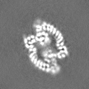
ジャーナル: Nat Struct Mol Biol / 年: 2024 タイトル: Structural mechanisms of autoinhibition and substrate recognition by the ubiquitin ligase HACE1. 著者: Jonas Düring / Madita Wolter / Julia J Toplak / Camilo Torres / Olexandr Dybkov / Thornton J Fokkens / Katherine E Bohnsack / Henning Urlaub / Wieland Steinchen / Christian Dienemann / Sonja Lorenz / 要旨: Ubiquitin ligases (E3s) are pivotal specificity determinants in the ubiquitin system by selecting substrates and decorating them with distinct ubiquitin signals. However, structure determination of ...Ubiquitin ligases (E3s) are pivotal specificity determinants in the ubiquitin system by selecting substrates and decorating them with distinct ubiquitin signals. However, structure determination of the underlying, specific E3-substrate complexes has proven challenging owing to their transient nature. In particular, it is incompletely understood how members of the catalytic cysteine-driven class of HECT-type ligases (HECTs) position substrate proteins for modification. Here, we report a cryogenic electron microscopy (cryo-EM) structure of the full-length human HECT HACE1, along with solution-based conformational analyses by small-angle X-ray scattering and hydrogen-deuterium exchange mass spectrometry. Structure-based functional analyses in vitro and in cells reveal that the activity of HACE1 is stringently regulated by dimerization-induced autoinhibition. The inhibition occurs at the first step of the catalytic cycle and is thus substrate-independent. We use mechanism-based chemical crosslinking to reconstitute a complex of activated, monomeric HACE1 with its major substrate, RAC1, determine its structure by cryo-EM and validate the binding mode by solution-based analyses. Our findings explain how HACE1 achieves selectivity in ubiquitinating the active, GTP-loaded state of RAC1 and establish a framework for interpreting mutational alterations of the HACE1-RAC1 interplay in disease. More broadly, this work illuminates central unexplored aspects in the architecture, conformational dynamics, regulation and specificity of full-length HECTs.
 ムービー
ムービー コントローラー


 データを開く
 基本情報
基本情報
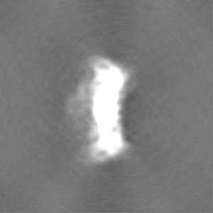
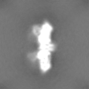
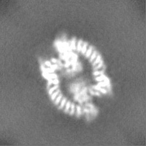
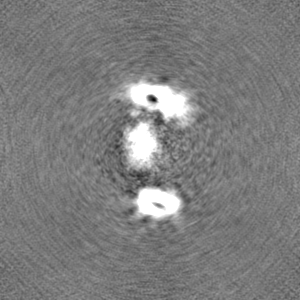
 マップデータ
 試料
試料 キーワード
キーワード 機能・相同性情報
 Homo sapiens (ヒト)
 データ登録者
 ドイツ, European Union, 2件
  引用
引用 構造の表示
構造の表示
 ダウンロードとリンク
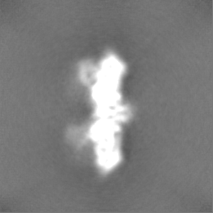
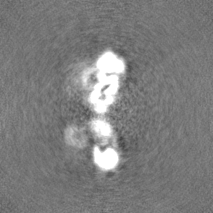
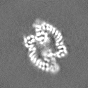
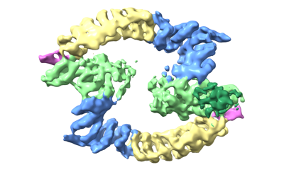 emd_17994.png
 http://ftp.pdbj.org/pub/emdb/structures/EMD-17994

 Z
Z Y
Y X
X


 試料の構成要素
 解析
解析 電子顕微鏡法
 FIELD EMISSION GUN