 Movie
Movie Controller

[English] 日本語
 Yorodumi
- EMDB-1736: Pyruvate carboxylase from S. aureus after addition of Acetyl-CoA -
+ Open data

- Basic information
| Entry | Database: EMDB / ID: EMD-1736 | |||||||||
|---|---|---|---|---|---|---|---|---|---|---|
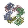
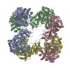
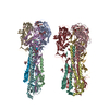
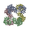
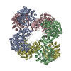
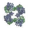
| Title | Pyruvate carboxylase from S. aureus after addition of Acetyl-CoA | |||||||||
 Map data Map data | Electron density map of pyruvate carboxylate after addition of acetyl-CoA | |||||||||
 Sample Sample |
| |||||||||
 Keywords Keywords | Pyruvate carboxylase / biotin-dependent carboxylase / acetyl-CoA / multifunctional / pyruvate / oxaloacetate / EC 6.4.1.1 | |||||||||
| Biological species |   Staphylococcus aureus (bacteria) Staphylococcus aureus (bacteria) | |||||||||
| Method | single particle reconstruction / cryo EM / Resolution: 13.4 Å | |||||||||
 Authors Authors | Lasso G / Yu LPC / Gil D / Xiang S / Tong L / Valle M | |||||||||
 Citation Citation | Journal: Structure / Year: 2010 Title: Cryo-EM analysis reveals new insights into the mechanism of action of pyruvate carboxylase. Authors: Gorka Lasso / Linda P C Yu / David Gil / Song Xiang / Liang Tong / Mikel Valle /  Abstract: Pyruvate carboxylase (PC) is a conserved multifunctional enzyme linked to important metabolic diseases. PC homotetramer is arranged in two layers with two opposing monomers per layer. Cryo-EM ...Pyruvate carboxylase (PC) is a conserved multifunctional enzyme linked to important metabolic diseases. PC homotetramer is arranged in two layers with two opposing monomers per layer. Cryo-EM explores the conformational variability of PC in the presence of different substrates. The results demonstrate that the biotin-carboxyl carrier protein (BCCP) domain localizes near the biotin carboxylase (BC) domain of its own monomer and travels to the carboxyltransferase (CT) domain of the opposite monomer. All density maps show noticeable conformational differences between layers, mainly for the BCCP and BC domains. This asymmetry may be indicative of a coordination mechanism where monomers from different layers catalyze the BC and CT reactions consecutively. A conformational change of the PC tetramerization (PT) domain suggests a new functional role in communication. A long-range communication pathway between subunits in different layers, via interacting PT-PT and BC-BC domains, may be responsible for the cooperativity of PC from Staphylococcus aureus. | |||||||||
| History |
|
- Structure visualization
| Movie |
 Movie viewer Movie viewer |
|---|---|
| Structure viewer | EM map: SurfViewMolmilJmol/JSmol |
| Supplemental images |

- Downloads & links
-EMDB archive
| Map data | emd_1736.map.gz | 5.7 MB | EMDB map data format | |
|---|---|---|---|---|
| Header (meta data) | emd-1736-v30.xmlemd-1736.xml | 9.4 KB 9.4 KB | Display Display | EMDB header |
| Images | 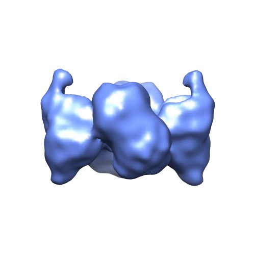 EMD1736.jpg EMD1736.jpg | 19.8 KB | ||
| Archive directory |  http://ftp.pdbj.org/pub/emdb/structures/EMD-1736ftp://ftp.pdbj.org/pub/emdb/structures/EMD-1736 http://ftp.pdbj.org/pub/emdb/structures/EMD-1736ftp://ftp.pdbj.org/pub/emdb/structures/EMD-1736 | HTTPS FTP |
-Validation report
| Summary document | emd_1736_validation.pdf.gz | 216.7 KB | Display | EMDB validaton report |
|---|---|---|---|---|
| Full document | emd_1736_full_validation.pdf.gz | 215.8 KB | Display | |
| Data in XML | emd_1736_validation.xml.gz | 5.6 KB | Display | |
| Arichive directory | https://ftp.pdbj.org/pub/emdb/validation_reports/EMD-1736ftp://ftp.pdbj.org/pub/emdb/validation_reports/EMD-1736 | HTTPS FTP |
-Related structure data
| Related structure data |  1737C  1738C  1741C  1742C 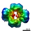 1743C  1744C C: citing same article ( |
|---|---|
| Similar structure data |

-Links
| EMDB pages | EMDB (EBI/PDBe) / EMDataResource |
|---|
-Map
| File | Download / File: emd_1736.map.gz / Format: CCP4 / Size: 6.1 MB / Type: IMAGE STORED AS FLOATING POINT NUMBER (4 BYTES) | ||||||||||||||||||||||||||||||||||||||||||||||||||||||||||||||||||||
|---|---|---|---|---|---|---|---|---|---|---|---|---|---|---|---|---|---|---|---|---|---|---|---|---|---|---|---|---|---|---|---|---|---|---|---|---|---|---|---|---|---|---|---|---|---|---|---|---|---|---|---|---|---|---|---|---|---|---|---|---|---|---|---|---|---|---|---|---|---|
| Annotation | Electron density map of pyruvate carboxylate after addition of acetyl-CoA | ||||||||||||||||||||||||||||||||||||||||||||||||||||||||||||||||||||
| Voxel size | X=Y=Z: 2.82 Å | ||||||||||||||||||||||||||||||||||||||||||||||||||||||||||||||||||||
| Density |
| ||||||||||||||||||||||||||||||||||||||||||||||||||||||||||||||||||||
| Symmetry | Space group: 1 | ||||||||||||||||||||||||||||||||||||||||||||||||||||||||||||||||||||
| Details | EMDB XML:
CCP4 map header:
| ||||||||||||||||||||||||||||||||||||||||||||||||||||||||||||||||||||
-Supplemental data
- Sample components
-Entire : Pyruvate carboxylase from S. aureus after addition of Acetyl-CoA
| Entire | Name: Pyruvate carboxylase from S. aureus after addition of Acetyl-CoA |
|---|---|
| Components |
|
-Supramolecule #1000: Pyruvate carboxylase from S. aureus after addition of Acetyl-CoA
| Supramolecule | Name: Pyruvate carboxylase from S. aureus after addition of Acetyl-CoA type: sample / ID: 1000 / Oligomeric state: Homotetramer / Number unique components: 1 |
|---|---|
| Molecular weight | Experimental: 520 KDa / Theoretical: 520 KDa |
-Macromolecule #1: Pyruvate carboxylase
| Macromolecule | Name: Pyruvate carboxylase / type: protein_or_peptide / ID: 1 / Name.synonym: PC / Oligomeric state: Homotetramer / Recombinant expression: Yes |
|---|---|
| Source (natural) | Organism: Staphylococcus aureus (bacteria) |
| Molecular weight | Theoretical: 520 KDa |
| Recombinant expression | Organism: Escherichia coli BL21 Star / Recombinant plasmid: pET28a |
-Experimental details
-Structure determination
| Method | cryo EM |
|---|---|
 Processing Processing | single particle reconstruction |
| Aggregation state | particle |
-Sample preparation
| Concentration | 0.1 mg/mL |
|---|---|
| Buffer | pH: 7.5 / Details: 20 mM Tris-HCL, 2mM NaCl, 2 mM DTT, 2mM acetyl-CoA |
| Grid | Details: quantifoil R2/2, 100 holey carbon films, Cu 200 mesh |
| Vitrification | Cryogen name: ETHANE / Chamber humidity: 100 % / Chamber temperature: 277 K / Instrument: OTHER / Details: Vitrification instrument: Vitrobot (FEI) / Timed resolved state: 45 sec / Method: Blot for 1.5 seconds |
- Electron microscopy
| Microscope | JEOL 2200FS |
|---|---|
| Temperature | Average: 99 K |
| Specialist optics | Energy filter - Name: Omega |
| Image recording | Category: FILM / Film or detector model: KODAK SO-163 FILM / Digitization - Scanner: ZEISS SCAI / Digitization - Sampling interval: 2.82 µm / Number real images: 88 / Average electron dose: 11 e/Å2 |
| Electron beam | Acceleration voltage: 200 kV / Electron source:  FIELD EMISSION GUN FIELD EMISSION GUN |
| Electron optics | Illumination mode: FLOOD BEAM / Imaging mode: BRIGHT FIELD / Cs: 2.0 mm / Nominal defocus max: 10.0 µm / Nominal defocus min: 2.32 µm / Nominal magnification: 50000 |
| Sample stage | Specimen holder: Single tilt cryoholder / Specimen holder model: GATAN LIQUID NITROGEN |
-Image processing
| Details | The particles were selected using a semi-automated procedure in spiderspire |
|---|---|
| CTF correction | Details: By defocus groups (Wiener filter) |
| Final reconstruction | Applied symmetry - Point group: C2 (2 fold cyclic) / Algorithm: OTHER / Resolution.type: BY AUTHOR / Resolution: 13.4 Å / Resolution method: OTHER / Software - Name: Spider / Number images used: 22258 |
| Final angle assignment | Details: Spider |
