Movie
Movie Controller

[English] 日本語
 Yorodumi
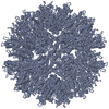
- EMDB-1710: Cryo-EM 3D model of the icosahedral particle composed of Rous sar... -
+ Open data

- Basic information
| Entry | Database: EMDB / ID: EMD-1710 | |||||||||
|---|---|---|---|---|---|---|---|---|---|---|
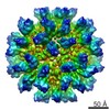
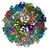
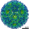
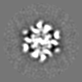
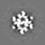
| Title | Cryo-EM 3D model of the icosahedral particle composed of Rous sarcoma virus capsid protein pentamers | |||||||||
 Map data Map data | This is a 3D reconstruction of the icosahedral particle composed of Rous sarcoma virus capsid protein pentamers | |||||||||
 Sample Sample |
| |||||||||
| Function / homology |  Function and homology information Function and homology informationhost cell nucleoplasm / viral procapsid maturation / host cell nucleolus / Hydrolases; Acting on peptide bonds (peptidases); Aspartic endopeptidases / viral capsid / structural constituent of virion / aspartic-type endopeptidase activity / nucleic acid binding / viral translational frameshifting / host cell plasma membrane ...host cell nucleoplasm / viral procapsid maturation / host cell nucleolus / Hydrolases; Acting on peptide bonds (peptidases); Aspartic endopeptidases / viral capsid / structural constituent of virion / aspartic-type endopeptidase activity / nucleic acid binding / viral translational frameshifting / host cell plasma membrane / proteolysis / zinc ion binding Similarity search - Function | |||||||||
| Biological species |  Rous sarcoma virus - Prague C Rous sarcoma virus - Prague C | |||||||||
| Method | single particle reconstruction / cryo EM / Resolution: 18.3 Å | |||||||||
 Authors Authors | Hyun JK / Radjainia M / Kingston RL / Mitra AK | |||||||||
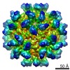
 Citation Citation | Journal: J Biol Chem / Year: 2010 Title: Proton-driven assembly of the Rous Sarcoma virus capsid protein results in the formation of icosahedral particles. Authors: Jae-Kyung Hyun / Mazdak Radjainia / Richard L Kingston / Alok K Mitra /  Abstract: In a mature and infectious retroviral particle, the capsid protein (CA) forms a shell surrounding the genomic RNA and the replicative machinery of the virus. The irregular nature of this capsid shell ...In a mature and infectious retroviral particle, the capsid protein (CA) forms a shell surrounding the genomic RNA and the replicative machinery of the virus. The irregular nature of this capsid shell precludes direct atomic resolution structural analysis. CA hexamers and pentamers are the fundamental building blocks of the capsid, however the pentameric state, in particular, remains poorly characterized. We have developed an efficient in vitro protocol for studying the assembly of Rous sarcoma virus (RSV) CA that involves mild acidification and produces structures modeling the authentic viral capsid. These structures include regular spherical particles with T = 1 icosahedral symmetry, built from CA pentamers alone. These particles were subject to cryoelectron microscopy (cryo-EM) and image processing, and a pseudo-atomic model of the icosahedron was created by docking atomic structures of the constituent CA domains into the cryo-EM-derived three-dimensional density map. The N-terminal domain (NTD) of CA forms pentameric turrets, which decorate the surface of the icosahedron, while the C-terminal domain (CTD) of CA is positioned underneath, linking the pentamers. Biophysical analysis of the icosahedral particle preparation reveals that CA monomers and icosahedra are the only detectable species and that these exist in reversible equilibrium at pH 5. These same acidic conditions are known to promote formation of a RSV CA CTD dimer, present within the icosahedral particle, which facilitates capsid assembly. The results are consistent with a model in which RSV CA assembly is a nucleation-limited process driven by very weak protein-protein interactions. | |||||||||
| History |
|
- Structure visualization
| Movie |
Movie viewer |
|---|---|
| Structure viewer | EM map: SurfViewMolmilJmol/JSmol |
| Supplemental images |
 UCSF Chimera
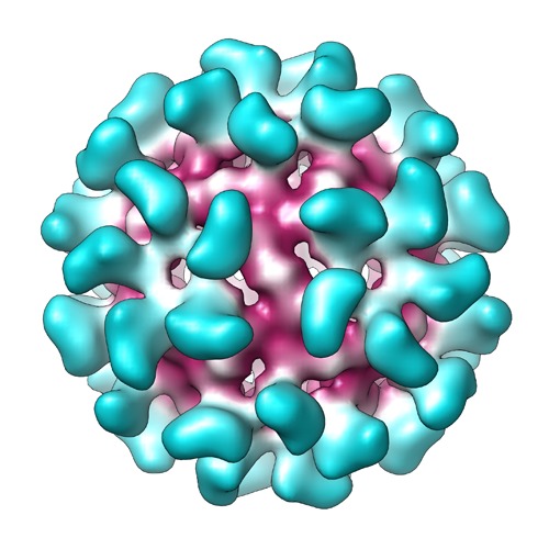
- Downloads & links
-EMDB archive
| Map data | emd_1710.map.gz | 7.4 MB | EMDB map data format | |
|---|---|---|---|---|
| Header (meta data) | emd-1710-v30.xmlemd-1710.xml | 10.7 KB 10.7 KB | Display Display | EMDB header |
| Images | map_EMD-1710.tif | 758.4 KB | ||
| Archive directory |  http://ftp.pdbj.org/pub/emdb/structures/EMD-1710ftp://ftp.pdbj.org/pub/emdb/structures/EMD-1710 http://ftp.pdbj.org/pub/emdb/structures/EMD-1710ftp://ftp.pdbj.org/pub/emdb/structures/EMD-1710 | HTTPS FTP |
-Related structure data
| Related structure data |  2x8qMC M: atomic model generated by this map C: citing same article ( |
|---|---|
| Similar structure data |

-Links
| EMDB pages | EMDB (EBI/PDBe) / EMDataResource |
|---|
-Map
| File | Download / File: emd_1710.map.gz / Format: CCP4 / Size: 15.3 MB / Type: IMAGE STORED AS FLOATING POINT NUMBER (4 BYTES) | ||||||||||||||||||||||||||||||||||||||||||||||||||||||||||||||||||||
|---|---|---|---|---|---|---|---|---|---|---|---|---|---|---|---|---|---|---|---|---|---|---|---|---|---|---|---|---|---|---|---|---|---|---|---|---|---|---|---|---|---|---|---|---|---|---|---|---|---|---|---|---|---|---|---|---|---|---|---|---|---|---|---|---|---|---|---|---|---|
| Annotation | This is a 3D reconstruction of the icosahedral particle composed of Rous sarcoma virus capsid protein pentamers | ||||||||||||||||||||||||||||||||||||||||||||||||||||||||||||||||||||
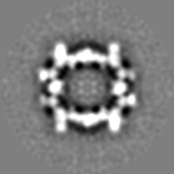
| Projections & slices | Image control
Images are generated by Spider. | ||||||||||||||||||||||||||||||||||||||||||||||||||||||||||||||||||||
| Voxel size | X=Y=Z: 2.5 Å | ||||||||||||||||||||||||||||||||||||||||||||||||||||||||||||||||||||
| Density |
| ||||||||||||||||||||||||||||||||||||||||||||||||||||||||||||||||||||
| Symmetry | Space group: 1 | ||||||||||||||||||||||||||||||||||||||||||||||||||||||||||||||||||||
| Details | EMDB XML:
CCP4 map header:
| ||||||||||||||||||||||||||||||||||||||||||||||||||||||||||||||||||||
 Z (Sec.)
 Y (Row.)
 X (Col.)


-Supplemental data
- Sample components
-Entire : Icosahedral particles composed of Rous sarcoma virus capsid protein
| Entire | Name: Icosahedral particles composed of Rous sarcoma virus capsid protein |
|---|---|
| Components |
|
-Supramolecule #1000: Icosahedral particles composed of Rous sarcoma virus capsid protein
| Supramolecule | Name: Icosahedral particles composed of Rous sarcoma virus capsid protein type: sample / ID: 1000 Details: The icosahedral particles were assembled in vitro, by transferring recombinant Rous sarcoma virus capsid protein monomers into high salt, mildly acidic buffer Oligomeric state: Icosahedral particle containing 12 CA pentamers (i.e. 60 monomers) Number unique components: 1 |
|---|---|
| Molecular weight | Theoretical: 1.5 MDa |
-Macromolecule #1: Capsid protein p27
| Macromolecule | Name: Capsid protein p27 / type: protein_or_peptide / ID: 1 / Name.synonym: Capsid protein p27 / Number of copies: 60 Oligomeric state: Icosahedral particle composed of 12 protein pentamers Recombinant expression: Yes |
|---|---|
| Source (natural) | Organism: Rous sarcoma virus - Prague C |
| Molecular weight | Theoretical: 1.53 MDa |
| Recombinant expression | Organism:  |
-Experimental details
-Structure determination
| Method | cryo EM |
|---|---|
 Processing Processing | single particle reconstruction |
| Aggregation state | particle |
-Sample preparation
| Concentration | 1.2 mg/mL |
|---|---|
| Buffer | pH: 5 Details: 0.1M citric acid, 5mM MOPS/KOH, 725mM NaCl, 0.25mM Na azide, 0.125mM TCEP-HCl |
| Grid | Details: Holey carbon 400 mesh copper grid |
| Vitrification | Cryogen name: ETHANE / Chamber humidity: 90 % / Chamber temperature: 85 K / Instrument: FEI VITROBOT MARK IV / Details: Vitrification instrument: Vitrobot Mark IV / Method: Blot for 5 seconds before plunging |
- Electron microscopy
| Microscope | FEI TECNAI 12 |
|---|---|
| Temperature | Average: 103 K |
| Alignment procedure | Legacy - Astigmatism: Objective lens astigmatism was corrected at 60,000 - 140,000 times magnification using live fft |
| Image recording | Category: FILM / Film or detector model: KODAK SO-163 FILM / Digitization - Scanner: NIKON SUPER COOLSCAN 9000 / Digitization - Sampling interval: 10.5 µm / Number real images: 21 / Average electron dose: 18 e/Å2 / Bits/pixel: 8 |
| Electron beam | Acceleration voltage: 120 kV / Electron source: LAB6 |
| Electron optics | Illumination mode: FLOOD BEAM / Imaging mode: BRIGHT FIELD / Cs: 2.0 mm / Nominal defocus max: 3.0 µm / Nominal defocus min: 0.8 µm / Nominal magnification: 42000 |
| Sample stage | Specimen holder: Side entry liquid nitrogen-cooled cryo specimen holder Specimen holder model: GATAN LIQUID NITROGEN |
-Image processing
| CTF correction | Details: Each micrograph |
|---|---|
| Final reconstruction | Applied symmetry - Point group: I (icosahedral) / Algorithm: OTHER / Resolution.type: BY AUTHOR / Resolution: 18.3 Å / Resolution method: FSC 0.5 CUT-OFF / Software - Name: Bsoft, PFT2, EM3DR2 Details: The digitized micrographs were processed using Bsoft. Orientation and origin search of the particles and 3D reconstruction were performed using PFT2 and EM3DR, respectively Number images used: 1310 |
-Atomic model buiding 1
| Initial model | (PDB ID: , ) |
|---|---|
| Software | Name: Chimera,Sculptor |
| Details | Protocol: Rigid body. The domain structures were manually fitted into the 3D reconstruction, and then the fitting was refined using Sculptor |
| Refinement | Space: RECIPROCAL / Protocol: RIGID BODY FIT / Target criteria: Cross-correlation |
| Output model | PDB-2x8q: |