- EMDB-16786: Human apoferritin after 561 nm laser exposure -
+
データを開く
IDまたはキーワード:
読み込み中...
-
基本情報
登録情報
データベース: EMDB / ID: EMD-16786
タイトル
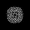
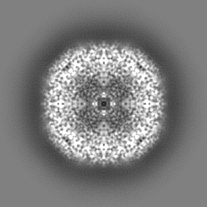
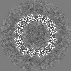
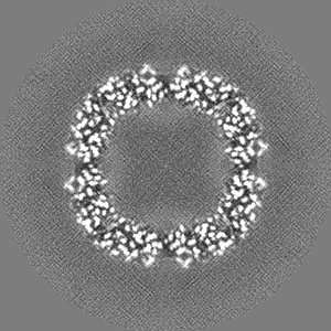
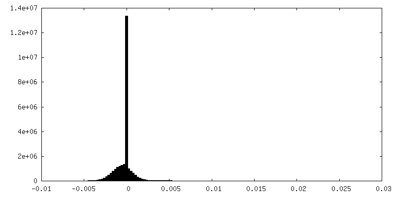
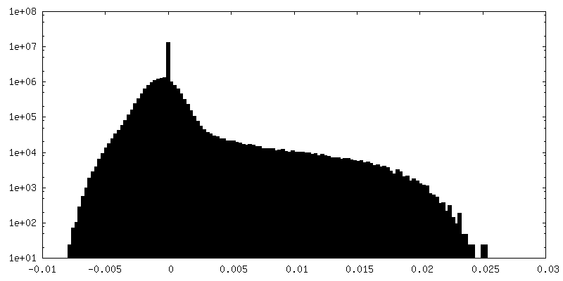
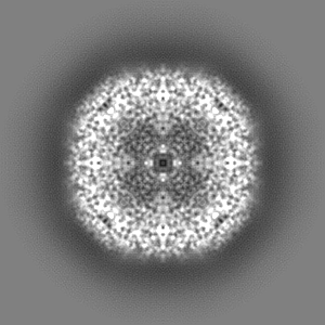
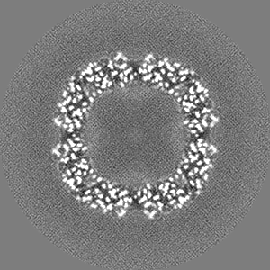
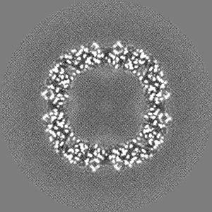
Human apoferritin after 561 nm laser exposure
マップデータ
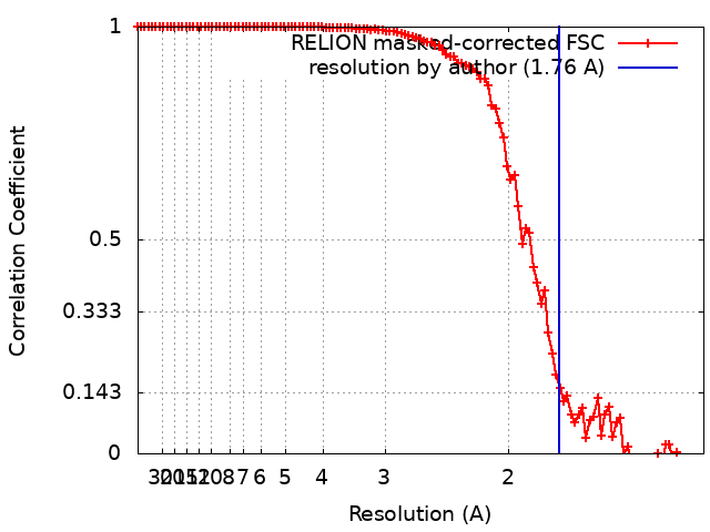
Map of human apoferritin acquired after illumination with 561 nm laser light (3.9e6 photons per sq. A).
試料
細胞器官・細胞要素: Apoferritin
タンパク質・ペプチド: Ferritin heavy chain, N-terminally processed
リガンド: SODIUM ION
リガンド: MAGNESIUM ION
リガンド: water
キーワード
Iron binding protein / METAL BINDING PROTEIN
機能・相同性
機能・相同性情報
iron ion sequestering activity / : / autolysosome / Scavenging by Class A Receptors / Golgi Associated Vesicle Biogenesis / ferroxidase / intracellular sequestering of iron ion / ferroxidase activity / negative regulation of fibroblast proliferation / ferric iron binding ...iron ion sequestering activity / : / autolysosome / Scavenging by Class A Receptors / Golgi Associated Vesicle Biogenesis / ferroxidase / intracellular sequestering of iron ion / ferroxidase activity / negative regulation of fibroblast proliferation / ferric iron binding / Iron uptake and transport / ferrous iron binding / tertiary granule lumen / iron ion transport / intracellular iron ion homeostasis / ficolin-1-rich granule lumen / iron ion binding / immune response / negative regulation of cell population proliferation / Neutrophil degranulation / extracellular exosome / extracellular region / identical protein binding / membrane / nucleus / cytoplasm / cytosol 類似検索 - 分子機能
Netherlands Organisation for Scientific Research (NWO)
VI.Vidi.193.014
オランダ
引用
ジャーナル: J Struct Biol / 年: 2023 タイトル: Super-resolution fluorescence imaging of cryosamples does not limit achievable resolution in cryoEM. 著者: Mart G F Last / Willem E M Noteborn / Lenard M Voortman / Thomas H Sharp / 要旨: Correlated super-resolution cryo-fluorescence and cryo-electron microscopy (cryoEM) has been gaining popularity as a method to investigate biological samples with high resolution and specificity. A ...Correlated super-resolution cryo-fluorescence and cryo-electron microscopy (cryoEM) has been gaining popularity as a method to investigate biological samples with high resolution and specificity. A concern in this combined method (called SR-cryoCLEM), however, is whether and how fluorescence imaging prior to cryoEM acquisition is detrimental to sample integrity. In this report, we investigated the effect of high-dose laser light (405, 488, and 561 nm) irradiation on apoferritin samples prepared for cryoEM with excitation wavelengths commonly used in fluorescence microscopy, and compared these samples to controls that were kept in the dark. We found that laser illumination, of equal duration and intensity as used in cryo-single molecule localization microscopy (cryoSMLM) and in the presence of high concentrations of fluorescent protein, did not affect the achievable resolution in cryoEM, with final reconstructions reaching resolutions of ∼ 1.8 Å regardless of the laser illumination. The finding that super-resolution fluorescence imaging of cryosamples prior to cryoEM data acquisition does not limit the achievable resolution suggests that super-resolution cryo-fluorescence microscopy and in situ structural biology using cryoEM are entirely compatible.
 ムービー
ムービー コントローラー


 データを開く
 基本情報
基本情報
 マップデータ
 試料
試料 キーワード
キーワード 機能・相同性情報
 Homo sapiens (ヒト)
 データ登録者
 オランダ, 1件
  引用
引用 構造の表示
構造の表示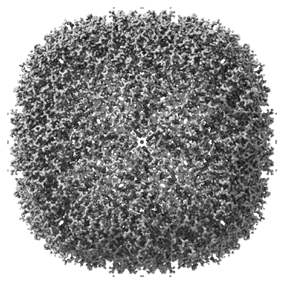
 ダウンロードとリンク
 emd_16786.png
 http://ftp.pdbj.org/pub/emdb/structures/EMD-16786


 Z (Sec.)
 Y (Row.)
 X (Col.)


 試料の構成要素

 解析
解析 電子顕微鏡法
 FIELD EMISSION GUN