 Movie
Movie Controller


+ Open data

- Basic information
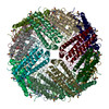
| Entry | Database: PDB / ID: 8cpu | ||||||
|---|---|---|---|---|---|---|---|
| Title | Human apoferritin after 561 nm laser exposure | ||||||
 Components Components | Ferritin heavy chain, N-terminally processed | ||||||
 Keywords Keywords | METAL BINDING PROTEIN / Iron binding protein | ||||||
| Function / homology |  Function and homology information Function and homology informationiron ion sequestering activity / ferritin complex / Scavenging by Class A Receptors / Golgi Associated Vesicle Biogenesis / ferroxidase / autolysosome / negative regulation of ferroptosis / ferroxidase activity / negative regulation of fibroblast proliferation / ferric iron binding ...iron ion sequestering activity / ferritin complex / Scavenging by Class A Receptors / Golgi Associated Vesicle Biogenesis / ferroxidase / autolysosome / negative regulation of ferroptosis / ferroxidase activity / negative regulation of fibroblast proliferation / ferric iron binding / autophagosome / iron ion transport / ferrous iron binding / Iron uptake and transport / tertiary granule lumen / ficolin-1-rich granule lumen / intracellular iron ion homeostasis / immune response / iron ion binding / negative regulation of cell population proliferation / Neutrophil degranulation / extracellular exosome / extracellular region / identical protein binding / nucleus / cytosol / cytoplasm Similarity search - Function | ||||||
| Biological species |  Homo sapiens (human) Homo sapiens (human) | ||||||
| Method | ELECTRON MICROSCOPY / single particle reconstruction / cryo EM / Resolution: 1.76 Å | ||||||
 Authors Authors | Last, M.G.F. / Noteborn, W.E.M. / Sharp, T.H. | ||||||
| Funding support |  Netherlands, 1items Netherlands, 1items
| ||||||
 Citation Citation | Journal: J Struct Biol / Year: 2023 Title: Super-resolution fluorescence imaging of cryosamples does not limit achievable resolution in cryoEM. Authors: Mart G F Last / Willem E M Noteborn / Lenard M Voortman / Thomas H Sharp / Abstract: Correlated super-resolution cryo-fluorescence and cryo-electron microscopy (cryoEM) has been gaining popularity as a method to investigate biological samples with high resolution and specificity. A ...Correlated super-resolution cryo-fluorescence and cryo-electron microscopy (cryoEM) has been gaining popularity as a method to investigate biological samples with high resolution and specificity. A concern in this combined method (called SR-cryoCLEM), however, is whether and how fluorescence imaging prior to cryoEM acquisition is detrimental to sample integrity. In this report, we investigated the effect of high-dose laser light (405, 488, and 561 nm) irradiation on apoferritin samples prepared for cryoEM with excitation wavelengths commonly used in fluorescence microscopy, and compared these samples to controls that were kept in the dark. We found that laser illumination, of equal duration and intensity as used in cryo-single molecule localization microscopy (cryoSMLM) and in the presence of high concentrations of fluorescent protein, did not affect the achievable resolution in cryoEM, with final reconstructions reaching resolutions of ∼ 1.8 Å regardless of the laser illumination. The finding that super-resolution fluorescence imaging of cryosamples prior to cryoEM data acquisition does not limit the achievable resolution suggests that super-resolution cryo-fluorescence microscopy and in situ structural biology using cryoEM are entirely compatible. | ||||||
| History |
|
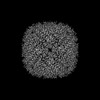
- Structure visualization
| Structure viewer | Molecule: MolmilJmol/JSmol |
|---|
- Downloads & links
-Download
| PDBx/mmCIF format | 8cpu.cif.gz | 743.1 KB | Display | PDBx/mmCIF format |
|---|---|---|---|---|
| PDB format | pdb8cpu.ent.gz | 602.6 KB | Display | PDB format |
| PDBx/mmJSON format | 8cpu.json.gz | Tree view | PDBx/mmJSON format | |
| Others |  Other downloads Other downloads |
-Validation report
| Arichive directory | https://data.pdbj.org/pub/pdb/validation_reports/cp/8cpuftp://data.pdbj.org/pub/pdb/validation_reports/cp/8cpu | HTTPS FTP |
|---|
-Related structure data
| Related structure data | 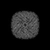 16786MC  8cpmC  8cpsC  8cptC  8cpvC  8cpwC  8cpxC M: map data used to model this data C: citing same article ( |
|---|---|
| Similar structure data |


-Links
 PDBj
PDBj


- Assembly
| Deposited unit | 
|
|---|---|
| 1 |
|
-Components
| #1: Protein | Mass: 20218.570 Da / Num. of mol.: 24 Source method: isolated from a genetically manipulated source Source: (gene. exp.) Homo sapiens (human) / Gene: FTH1, FTH, FTHL6, OK/SW-cl.84, PIG15 / Production host:  #2: Chemical | ChemComp-NA /   Mass: 22.990 Da / Num. of mol.: 24 / Source method: obtained synthetically / Formula: Na Mass: 22.990 Da / Num. of mol.: 24 / Source method: obtained synthetically / Formula: Na#3: Chemical | ChemComp-MG /   Mass: 24.305 Da / Num. of mol.: 14 / Source method: obtained synthetically / Formula: Mg Mass: 24.305 Da / Num. of mol.: 14 / Source method: obtained synthetically / Formula: Mg#4: Water | ChemComp-HOH / |  Mass: 18.015 Da / Num. of mol.: 1705 / Source method: isolated from a natural source / Formula: H2O Mass: 18.015 Da / Num. of mol.: 1705 / Source method: isolated from a natural source / Formula: H2OHas ligand of interest | N | |
|---|
-Experimental details
-Experiment
| Experiment | Method: ELECTRON MICROSCOPY |
|---|---|
| EM experiment | Aggregation state: PARTICLE / 3D reconstruction method: single particle reconstruction |
- Sample preparation
| Component | Name: Apoferritin / Type: ORGANELLE OR CELLULAR COMPONENT / Entity ID: #1 / Source: RECOMBINANT | |||||||||||||||
|---|---|---|---|---|---|---|---|---|---|---|---|---|---|---|---|---|
| Molecular weight | Value: 0.484 MDa / Experimental value: NO | |||||||||||||||
| Source (natural) | Organism: Homo sapiens (human) | |||||||||||||||
| Source (recombinant) | Organism: | |||||||||||||||
| Buffer solution | pH: 8 | |||||||||||||||
| Buffer component |
| |||||||||||||||
| Specimen | Conc.: 4 mg/ml / Embedding applied: NO / Shadowing applied: NO / Staining applied: NO / Vitrification applied: YES | |||||||||||||||
| Specimen support | Grid material: GOLD / Grid mesh size: 300 divisions/in. / Grid type: Quantifoil R1.2/1.3 | |||||||||||||||
| Vitrification | Instrument: FEI VITROBOT MARK IV / Cryogen name: ETHANE / Humidity: 100 % / Chamber temperature: 295 K |
- Electron microscopy imaging
| Experimental equipment |  Model: Titan Krios / Image courtesy: FEI Company |
|---|---|
| Microscopy | Model: TFS KRIOS |
| Electron gun | Electron source:  FIELD EMISSION GUN / Accelerating voltage: 300 kV / Illumination mode: FLOOD BEAM FIELD EMISSION GUN / Accelerating voltage: 300 kV / Illumination mode: FLOOD BEAM |
| Electron lens | Mode: BRIGHT FIELD / Nominal magnification: 130000 X / Nominal defocus max: 1500 nm / Nominal defocus min: 500 nm / Cs: 2.7 mm / C2 aperture diameter: 50 µm |
| Specimen holder | Cryogen: NITROGEN / Specimen holder model: FEI TITAN KRIOS AUTOGRID HOLDER |
| Image recording | Electron dose: 50 e/Å2 / Film or detector model: GATAN K3 BIOQUANTUM (6k x 4k) |
| EM imaging optics | Energyfilter name: GIF Bioquantum / Energyfilter slit width: 20 eV |
- Processing
| Software | Name: UCSF ChimeraX / Version: 1.4/v9 / Classification: model building / URL: https://www.rbvi.ucsf.edu/chimerax/ / Os: Windows / Type: package | ||||||||||||||||||||||||
|---|---|---|---|---|---|---|---|---|---|---|---|---|---|---|---|---|---|---|---|---|---|---|---|---|---|
| EM software |
| ||||||||||||||||||||||||
| CTF correction | Type: PHASE FLIPPING AND AMPLITUDE CORRECTION | ||||||||||||||||||||||||
| 3D reconstruction | Resolution: 1.76 Å / Resolution method: FSC 0.143 CUT-OFF / Num. of particles: 9700 / Symmetry type: POINT | ||||||||||||||||||||||||
| Atomic model building | PDB-ID: 6Z6U Accession code: 6Z6U / Source name: PDB / Type: experimental model |
