 Movie
Movie Controller

[English] 日本語
 Yorodumi
- EMDB-1661: The three-dimensional structure of a hepatitis C virus p7 ion cha... -
+ Open data

- Basic information
| Entry | Database: EMDB / ID: EMD-1661 | |||||||||
|---|---|---|---|---|---|---|---|---|---|---|
| Title | The three-dimensional structure of a hepatitis C virus p7 ion channel by electron microscopy | |||||||||
 Map data Map data | Top view of the EM density map of the HCV p7 channel | |||||||||
 Sample Sample |
| |||||||||
| Biological species |  Hepatitis C virus Hepatitis C virus | |||||||||
| Method | single particle reconstruction / negative staining / Resolution: 16.0 Å | |||||||||
 Authors Authors | Luik P / Chew C / Aittoniemi J / Chang J / Wentworth P / Dwek RA / Biggin PC / Venien-Bryan C / Zitzmann N | |||||||||
 Citation Citation | Journal: Proc Natl Acad Sci U S A / Year: 2009 Title: The 3-dimensional structure of a hepatitis C virus p7 ion channel by electron microscopy. Authors: Philipp Luik / Chee Chew / Jussi Aittoniemi / Jason Chang / Paul Wentworth / Raymond A Dwek / Philip C Biggin / Catherine Vénien-Bryan / Nicole Zitzmann /  Abstract: Infection with the hepatitis C virus (HCV) has a huge impact on global health putting more than 170 million people at risk of developing severe liver disease. The HCV encoded p7 ion channel is ...Infection with the hepatitis C virus (HCV) has a huge impact on global health putting more than 170 million people at risk of developing severe liver disease. The HCV encoded p7 ion channel is essential for the production of infectious viruses. Despite a growing body of functional data, little is known about the 3-dimensional (3D) structure of the channel. Here, we present the 3D structure of a full-length viroporin, the detergent-solubilized hexameric 42 kDa form of the HCV p7 ion channel, as determined by single-particle electron microscopy using the random conical tilting approach. The reconstruction of such a small protein complex was made possible by a combination of high-contrast staining, the symmetry, and the distinct structural features of the channel. The orientation of the p7 monomers within the density was established using immunolabeling with N and C termini specific F(ab) fragments. The density map at a resolution of approximately 16 A reveals a flower-shaped protein architecture with protruding petals oriented toward the ER lumen. This broadest part of the channel presents a comparatively large surface area providing potential interaction sites for cellular and virally encoded ER resident proteins. | |||||||||
| History |
|
- Structure visualization
| Movie |
 Movie viewer Movie viewer |
|---|---|
| Structure viewer | EM map: SurfViewMolmilJmol/JSmol |
| Supplemental images |
 UCSF Chimera
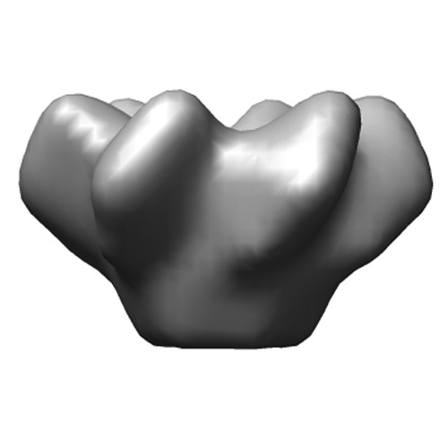
- Downloads & links
-EMDB archive
| Map data | emd_1661.map.gz | 968.9 KB | EMDB map data format | |
|---|---|---|---|---|
| Header (meta data) | emd-1661-v30.xmlemd-1661.xml | 7.2 KB 7.2 KB | Display Display | EMDB header |
| Images | Preview_EMD-1661.tif | 256.4 KB | ||
| Archive directory |  http://ftp.pdbj.org/pub/emdb/structures/EMD-1661ftp://ftp.pdbj.org/pub/emdb/structures/EMD-1661 http://ftp.pdbj.org/pub/emdb/structures/EMD-1661ftp://ftp.pdbj.org/pub/emdb/structures/EMD-1661 | HTTPS FTP |
-Validation report
| Summary document | emd_1661_validation.pdf.gz | 190.4 KB | Display | EMDB validaton report |
|---|---|---|---|---|
| Full document | emd_1661_full_validation.pdf.gz | 189.5 KB | Display | |
| Data in XML | emd_1661_validation.xml.gz | 4.8 KB | Display | |
| Arichive directory | https://ftp.pdbj.org/pub/emdb/validation_reports/EMD-1661ftp://ftp.pdbj.org/pub/emdb/validation_reports/EMD-1661 | HTTPS FTP |
-Related structure data
| Similar structure data |
|---|


-Links
| EMDB pages | EMDB (EBI/PDBe) / EMDataResource |
|---|
-Map
| File | Download / File: emd_1661.map.gz / Format: CCP4 / Size: 1001 KB / Type: IMAGE STORED AS FLOATING POINT NUMBER (4 BYTES) | ||||||||||||||||||||||||||||||||||||||||||||||||||||||||||||||||||||
|---|---|---|---|---|---|---|---|---|---|---|---|---|---|---|---|---|---|---|---|---|---|---|---|---|---|---|---|---|---|---|---|---|---|---|---|---|---|---|---|---|---|---|---|---|---|---|---|---|---|---|---|---|---|---|---|---|---|---|---|---|---|---|---|---|---|---|---|---|---|
| Annotation | Top view of the EM density map of the HCV p7 channel | ||||||||||||||||||||||||||||||||||||||||||||||||||||||||||||||||||||
| Projections & slices | Image control
Images are generated by Spider. | ||||||||||||||||||||||||||||||||||||||||||||||||||||||||||||||||||||
| Voxel size | X=Y=Z: 2.77 Å | ||||||||||||||||||||||||||||||||||||||||||||||||||||||||||||||||||||
| Density |
| ||||||||||||||||||||||||||||||||||||||||||||||||||||||||||||||||||||
| Symmetry | Space group: 1 | ||||||||||||||||||||||||||||||||||||||||||||||||||||||||||||||||||||
| Details | EMDB XML:
CCP4 map header:
| ||||||||||||||||||||||||||||||||||||||||||||||||||||||||||||||||||||
 Z (Sec.)
 Y (Row.)
 X (Col.)


-Supplemental data
- Sample components
-Entire : HCV p7 hexamer, JFH-1 strain, genotype 2a
| Entire | Name: HCV p7 hexamer, JFH-1 strain, genotype 2a |
|---|---|
| Components |
|
-Supramolecule #1000: HCV p7 hexamer, JFH-1 strain, genotype 2a
| Supramolecule | Name: HCV p7 hexamer, JFH-1 strain, genotype 2a / type: sample / ID: 1000 / Oligomeric state: Hexameric / Number unique components: 1 |
|---|
-Macromolecule #1: p7
| Macromolecule | Name: p7 / type: protein_or_peptide / ID: 1 / Name.synonym: p7 / Details: p7 solubilzed with DHPC / Recombinant expression: No |
|---|---|
| Source (natural) | Organism: Hepatitis C virus |
-Experimental details
-Structure determination
| Method | negative staining |
|---|---|
 Processing Processing | single particle reconstruction |
| Aggregation state | particle |
-Sample preparation
| Buffer | pH: 7 / Details: 100 mM NaCl, 50 mM DHPC, 25 mM Hepes, pH 7.0 |
|---|---|
| Staining | Type: NEGATIVE / Details: 2% (w/v) phosphotungstic acid (PTA) |
| Vitrification | Cryogen name: NONE / Instrument: OTHER |
- Electron microscopy
| Microscope | FEI/PHILIPS CM120T |
|---|---|
| Image recording | Category: FILM / Film or detector model: KODAK SO-163 FILM / Digitization - Scanner: NIKON SUPER COOLSCAN 9000 |
| Electron beam | Acceleration voltage: 120 kV / Electron source: LAB6 |
| Electron optics | Illumination mode: SPOT SCAN / Imaging mode: BRIGHT FIELD |
| Sample stage | Specimen holder: Eucentric / Specimen holder model: PHILIPS ROTATION HOLDER |
-Image processing
| Final reconstruction | Applied symmetry - Point group: C6 (6 fold cyclic) / Resolution.type: BY AUTHOR / Resolution: 16.0 Å / Resolution method: FSC 0.5 CUT-OFF |
|---|
