- EMDB-1430: Specific interaction between EF-G and RRF and its implication for... -
+
Open data
ID or keywords:
Loading...
-
Basic information
Entry
Database: EMDB / ID: EMD-1430
Title
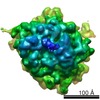
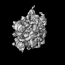
Specific interaction between EF-G and RRF and its implication for GTP-dependent ribosome splitting into subunits.
Map data
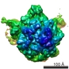
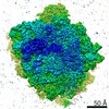
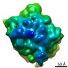
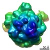
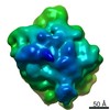
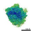
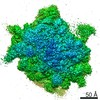
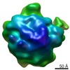
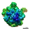
Cryo-EM map of E.coli 50S complex
Sample
Sample: 50S subunit with EF-G and RRF bound
Complex: 50S subunit
Protein or peptide: EF-G
Protein or peptide: RRF
Function / homology
Function and homology information
cytoplasmic translational termination / ribosome disassembly / guanosine tetraphosphate binding / stringent response / negative regulation of cytoplasmic translational initiation / ribosomal large subunit binding / translational elongation / translation elongation factor activity / translational termination / transcriptional attenuation ...cytoplasmic translational termination / ribosome disassembly / guanosine tetraphosphate binding / stringent response / negative regulation of cytoplasmic translational initiation / ribosomal large subunit binding / translational elongation / translation elongation factor activity / translational termination / transcriptional attenuation / endoribonuclease inhibitor activity / positive regulation of ribosome biogenesis / RNA-binding transcription regulator activity / negative regulation of cytoplasmic translation / DnaA-L2 complex / translation repressor activity / negative regulation of translational initiation / negative regulation of DNA-templated DNA replication initiation / mRNA regulatory element binding translation repressor activity / response to reactive oxygen species / cytosolic ribosome assembly / ribosome assembly / assembly of large subunit precursor of preribosome / translational initiation / regulation of cell growth / DNA-templated transcription termination / response to radiation / mRNA 5'-UTR binding / large ribosomal subunit / transferase activity / ribosome binding / 5S rRNA binding / ribosomal large subunit assembly / large ribosomal subunit rRNA binding / cytosolic large ribosomal subunit / Hydrolases; Acting on acid anhydrides; Acting on GTP to facilitate cellular and subcellular movement / cytoplasmic translation / tRNA binding / negative regulation of translation / rRNA binding / structural constituent of ribosome / ribosome / translation / response to antibiotic / negative regulation of DNA-templated transcription / mRNA binding / GTPase activity / GTP binding / DNA binding / RNA binding / zinc ion binding / cytoplasm / cytosol Similarity search - Function
Ribosome recycling factor / Ribosome recycling factor domain / RRF superfamily / Ribosome recycling factor / Translation elongation factor EFG/EF2 / : / Elongation factor G, domain III / EFG, domain V / Ribosomal protein L1, bacterial-type / Elongation Factor G, domain II ...Ribosome recycling factor / Ribosome recycling factor domain / RRF superfamily / Ribosome recycling factor / Translation elongation factor EFG/EF2 / : / Elongation factor G, domain III / EFG, domain V / Ribosomal protein L1, bacterial-type / Elongation Factor G, domain II / Elongation Factor G, domain III / Translation elongation factor EFG/EF2, domain IV / Elongation factor G, domain IV / Elongation factor G, domain IV / Elongation factor G C-terminus / Elongation factor EFG, domain V-like / Elongation factor G C-terminus / EF-G domain III/V-like / Tr-type G domain, conserved site / Translational (tr)-type guanine nucleotide-binding (G) domain signature. / Ribosomal protein L1, conserved site / Ribosomal protein L1 signature. / Ribosomal protein L1 / Ribosomal protein L1, 3-layer alpha/beta-sandwich / Translation elongation factor EFTu-like, domain 2 / Ribosomal protein L1-like / Ribosomal protein L1/ribosomal biogenesis protein / Ribosomal protein L1p/L10e family / Ribosomal protein L11, bacterial-type / Elongation factor Tu domain 2 / Ribosomal protein L25, short-form / Ribosomal protein L31 type A / Translational (tr)-type GTP-binding domain / Elongation factor Tu GTP binding domain / Translational (tr)-type guanine nucleotide-binding (G) domain profile. / Ribosomal protein L31 signature. / Ribosomal protein L11, conserved site / Ribosomal protein L11 signature. / Ribosomal protein L31 / Ribosomal protein L31 superfamily / Ribosomal protein L31 / Ribosomal protein L9 signature. / Ribosomal protein L16 signature 1. / Ribosomal protein L6, conserved site / Ribosomal protein L6 signature 1. / Ribosomal protein L9, bacteria/chloroplast / Ribosomal protein L9, C-terminal / Ribosomal protein L9, C-terminal domain / Ribosomal protein L21, conserved site / Ribosomal protein L21 signature. / : / Ribosomal protein L11, N-terminal / Ribosomal protein L9, C-terminal domain superfamily / Ribosomal protein L11, N-terminal domain / Ribosomal protein L11/L12 / Ribosomal protein L11, C-terminal / Ribosomal protein L11, C-terminal domain superfamily / Ribosomal protein L11/L12, N-terminal domain superfamily / Ribosomal protein L11/L12 / Ribosomal protein L11, RNA binding domain / Ribosomal protein L16 signature 2. / Ribosomal protein L16, conserved site / Ribosomal protein L17 signature. / Ribosomal L25p family / Ribosomal protein L25 / Ribosomal protein L36 signature. / Ribosomal protein L25/Gln-tRNA synthetase, N-terminal / Ribosomal protein L25/Gln-tRNA synthetase, anti-codon-binding domain superfamily / : / Ribosomal protein L33, conserved site / Ribosomal protein L33 signature. / Ribosomal protein L32p, bacterial type / Ribosomal protein L35, conserved site / Ribosomal protein L35 signature. / Ribosomal protein L9 / Ribosomal protein L9, N-terminal domain superfamily / Ribosomal protein L9, N-terminal / Ribosomal protein L9, N-terminal domain / Ribosomal protein L35, non-mitochondrial / Ribosomal protein L18, bacterial-type / : / Ribosomal protein L6, bacterial-type / Ribosomal protein L5, bacterial-type / Ribosomal protein L9/RNase H1, N-terminal / Ribosomal protein L19, conserved site / Ribosomal protein L19 signature. / : / Ribosomal protein L36 / Ribosomal protein L36 superfamily / Ribosomal protein L36 / Ribosomal protein L20 signature. / Ribosomal protein L34, conserved site / Ribosomal protein L34 signature. / Ribosomal protein L14P, bacterial-type / Ribosomal protein L27, conserved site / Ribosomal protein L27 signature. / Ribosomal protein L35 / Ribosomal protein L35 superfamily / Ribosomal protein L22, bacterial/chloroplast-type / Ribosomal protein L35 Similarity search - Domain/homology
Large ribosomal subunit protein uL15 / Elongation factor G / Large ribosomal subunit protein uL11 / Large ribosomal subunit protein bL19 / Large ribosomal subunit protein uL1 / Large ribosomal subunit protein bL20 / Large ribosomal subunit protein bL27 / Large ribosomal subunit protein uL29 / Large ribosomal subunit protein bL31 / Large ribosomal subunit protein bL32 ...Large ribosomal subunit protein uL15 / Elongation factor G / Large ribosomal subunit protein uL11 / Large ribosomal subunit protein bL19 / Large ribosomal subunit protein uL1 / Large ribosomal subunit protein bL20 / Large ribosomal subunit protein bL27 / Large ribosomal subunit protein uL29 / Large ribosomal subunit protein bL31 / Large ribosomal subunit protein bL32 / Large ribosomal subunit protein bL33 / Large ribosomal subunit protein bL34 / Large ribosomal subunit protein bL35 / Large ribosomal subunit protein bL36A / Large ribosomal subunit protein bL9 / Ribosome-recycling factor / Large ribosomal subunit protein uL13 / Large ribosomal subunit protein uL14 / Large ribosomal subunit protein uL16 / Large ribosomal subunit protein uL23 / Large ribosomal subunit protein bL17 / Large ribosomal subunit protein bL21 / Large ribosomal subunit protein uL30 / Large ribosomal subunit protein uL6 / Large ribosomal subunit protein uL18 / Large ribosomal subunit protein uL2 / Large ribosomal subunit protein uL3 / Large ribosomal subunit protein uL24 / Large ribosomal subunit protein uL4 / Large ribosomal subunit protein uL22 / Large ribosomal subunit protein uL5 / Large ribosomal subunit protein bL25 Similarity search - Component
Biological species
Escherichia coli (E. coli)
Method
single particle reconstruction / cryo EM / Resolution: 9.1 Å
Journal: J Mol Biol / Year: 2007 Title: Specific interaction between EF-G and RRF and its implication for GTP-dependent ribosome splitting into subunits. Authors: Ning Gao / Andrey V Zavialov / Måns Ehrenberg / Joachim Frank / Abstract: After termination of protein synthesis, the bacterial ribosome is split into its 30S and 50S subunits by the action of ribosome recycling factor (RRF) and elongation factor G (EF-G) in a guanosine 5'- ...After termination of protein synthesis, the bacterial ribosome is split into its 30S and 50S subunits by the action of ribosome recycling factor (RRF) and elongation factor G (EF-G) in a guanosine 5'-triphosphate (GTP)-hydrolysis-dependent manner. Based on a previous cryo-electron microscopy study of ribosomal complexes, we have proposed that the binding of EF-G to an RRF-containing posttermination ribosome triggers an interdomain rotation of RRF, which destabilizes two strong intersubunit bridges (B2a and B3) and, ultimately, separates the two subunits. Here, we present a 9-A (Fourier shell correlation cutoff of 0.5) cryo-electron microscopy map of a 50S x EF-G x guanosine 5'-[(betagamma)-imido]triphosphate x RRF complex and a quasi-atomic model derived from it, showing the interaction between EF-G and RRF on the 50S subunit in the presence of the noncleavable GTP analogue guanosine 5'-[(betagamma)-imido]triphosphate. The detailed information in this model and a comparative analysis of EF-G structures in various nucleotide- and ribosome-bound states show how rotation of the RRF head domain may be triggered by various domains of EF-G. For validation of our structural model, all known mutations in EF-G and RRF that relate to ribosome recycling have been taken into account. More importantly, our results indicate a substantial conformational change in the Switch I region of EF-G, suggesting that a conformational signal transduction mechanism, similar to that employed in transfer RNA translocation on the ribosome by EF-G, translates a large-scale movement of EF-G's domain IV, induced by GTP hydrolysis, into the domain rotation of RRF that eventually splits the ribosome into subunits.
History
Deposition
Sep 24, 2007
-
Header (metadata) release
Sep 24, 2007
-
Map release
Dec 12, 2007
-
Update
Nov 7, 2012
-
Current status
Nov 7, 2012
Processing site: PDBe / Status: Released
-
Structure visualization
Movie
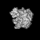
Surface view with section colored by density value
Category: FILM / Film or detector model: KODAK SO-163 FILM / Digitization - Scanner: ZEISS SCAI / Digitization - Sampling interval: 14 µm / Number real images: 259 / Average electron dose: 15 e/Å2
Tilt angle min
0
Tilt angle max
0
Electron beam
Acceleration voltage: 200 kV / Electron source: FIELD EMISSION GUN
In the structure databanks used in Yorodumi, some data are registered as the other names, "COVID-19 virus" and "2019-nCoV". Here are the details of the virus and the list of structure data.
Jan 31, 2019. EMDB accession codes are about to change! (news from PDBe EMDB page)
EMDB accession codes are about to change! (news from PDBe EMDB page)
The allocation of 4 digits for EMDB accession codes will soon come to an end. Whilst these codes will remain in use, new EMDB accession codes will include an additional digit and will expand incrementally as the available range of codes is exhausted. The current 4-digit format prefixed with “EMD-” (i.e. EMD-XXXX) will advance to a 5-digit format (i.e. EMD-XXXXX), and so on. It is currently estimated that the 4-digit codes will be depleted around Spring 2019, at which point the 5-digit format will come into force.
The EM Navigator/Yorodumi systems omit the EMD- prefix.
Related info.:Q: What is EMD? / ID/Accession-code notation in Yorodumi/EM Navigator
Yorodumi is a browser for structure data from EMDB, PDB, SASBDB, etc.
This page is also the successor to EM Navigator detail page, and also detail information page/front-end page for Omokage search.
The word "yorodu" (or yorozu) is an old Japanese word meaning "ten thousand". "mi" (miru) is to see.
Related info.:EMDB / PDB / SASBDB / Comparison of 3 databanks / Yorodumi Search / Aug 31, 2016. New EM Navigator & Yorodumi / Yorodumi Papers / Jmol/JSmol / Function and homology information / Changes in new EM Navigator and Yorodumi
 Movie
Movie Controller

 Yorodumi
 Open data
 Basic information
 Map data
 Sample
 Function and homology information
 Authors
 Citation
 Structure visualization
 Downloads & links
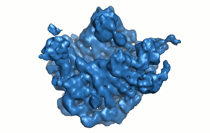 1430.gif
 http://ftp.pdbj.org/pub/emdb/structures/EMD-1430


 Z (Sec.)
 Y (Row.)
 X (Col.)


 Sample components
 Processing
 Electron microscopy
 FIELD EMISSION GUN
