Movie
Movie Controller


+ Open data

- Basic information
| Entry | Database: EMDB / ID: EMD-0372 | ||||||||||||||||||
|---|---|---|---|---|---|---|---|---|---|---|---|---|---|---|---|---|---|---|---|
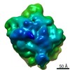
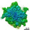
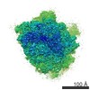
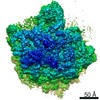
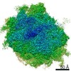
| Title | Cryo-EM structure of pre-Lsg1 (PL) pre-60S ribosomal subunit | ||||||||||||||||||
 Map data Map data | Cryo-EM structure of pre-Lsg1 (PL) pre-60S ribosomal subunit | ||||||||||||||||||
 Sample Sample |
| ||||||||||||||||||
 Keywords Keywords | ribosome biogenesis / Nmd3 / peptidyl transferase center / RIBOSOME | ||||||||||||||||||
| Function / homology |  Function and homology information Function and homology informationprotein tyrosine/serine/threonine phosphatase activity / ascospore wall assembly / preribosome binding / positive regulation of autophagosome assembly / ribophagy / maturation of 5.8S rRNA from tricistronic rRNA transcript (SSU-rRNA, 5.8S rRNA, LSU-rRNA) / cellular response to nitrogen starvation / pre-mRNA 5'-splice site binding / cytosolic large ribosomal subunit assembly / maturation of 5.8S rRNA ...protein tyrosine/serine/threonine phosphatase activity / ascospore wall assembly / preribosome binding / positive regulation of autophagosome assembly / ribophagy / maturation of 5.8S rRNA from tricistronic rRNA transcript (SSU-rRNA, 5.8S rRNA, LSU-rRNA) / cellular response to nitrogen starvation / pre-mRNA 5'-splice site binding / cytosolic large ribosomal subunit assembly / maturation of 5.8S rRNA / response to cycloheximide / cleavage in ITS2 between 5.8S rRNA and LSU-rRNA of tricistronic rRNA transcript (SSU-rRNA, 5.8S rRNA, LSU-rRNA) / SRP-dependent cotranslational protein targeting to membrane / ribosomal large subunit binding / GTP hydrolysis and joining of the 60S ribosomal subunit / negative regulation of mRNA splicing, via spliceosome / preribosome, large subunit precursor / Formation of a pool of free 40S subunits / Nonsense Mediated Decay (NMD) independent of the Exon Junction Complex (EJC) / Nonsense Mediated Decay (NMD) enhanced by the Exon Junction Complex (EJC) / L13a-mediated translational silencing of Ceruloplasmin expression / translational elongation / ribosomal large subunit export from nucleus / ribosomal subunit export from nucleus / regulation of translational fidelity / maturation of LSU-rRNA / protein-RNA complex assembly / protein-tyrosine-phosphatase / translation initiation factor activity / protein tyrosine phosphatase activity / ribosomal large subunit biogenesis / cytosolic ribosome assembly / maturation of LSU-rRNA from tricistronic rRNA transcript (SSU-rRNA, 5.8S rRNA, LSU-rRNA) / assembly of large subunit precursor of preribosome / meiotic cell cycle / macroautophagy / translational initiation / maintenance of translational fidelity / modification-dependent protein catabolic process / cytoplasmic stress granule / protein tag activity / rRNA processing / protein transport / ribosome biogenesis / 5S rRNA binding / ribosomal large subunit assembly / large ribosomal subunit rRNA binding / cytosolic large ribosomal subunit / protein-macromolecule adaptor activity / cytoplasmic translation / negative regulation of translation / rRNA binding / structural constituent of ribosome / protein ubiquitination / ribosome / translation / response to antibiotic / mRNA binding / ubiquitin protein ligase binding / nucleolus / RNA binding / zinc ion binding / nucleoplasm / nucleus / cytoplasm / cytosol Similarity search - Function | ||||||||||||||||||
| Biological species |  | ||||||||||||||||||
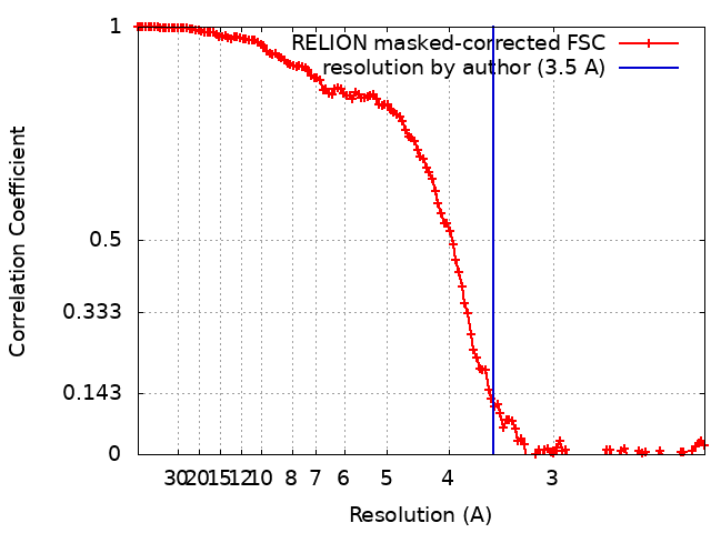
| Method | single particle reconstruction / cryo EM / Resolution: 3.5 Å | ||||||||||||||||||
 Authors Authors | Zhou Y / Musalgaonkar S | ||||||||||||||||||
| Funding support |  United States, 5 items United States, 5 items
| ||||||||||||||||||
 Citation Citation | Journal: Nat Commun / Year: 2019 Title: Tightly-orchestrated rearrangements govern catalytic center assembly of the ribosome. Authors: Yi Zhou / Sharmishtha Musalgaonkar / Arlen W Johnson / David W Taylor / Abstract: The catalytic activity of the ribosome is mediated by RNA, yet proteins are essential for the function of the peptidyl transferase center (PTC). In eukaryotes, final assembly of the PTC occurs in the ...The catalytic activity of the ribosome is mediated by RNA, yet proteins are essential for the function of the peptidyl transferase center (PTC). In eukaryotes, final assembly of the PTC occurs in the cytoplasm by insertion of the ribosomal protein Rpl10 (uL16). We determine structures of six intermediates in late nuclear and cytoplasmic maturation of the large subunit that reveal a tightly-choreographed sequence of protein and RNA rearrangements controlling the insertion of Rpl10. We also determine the structure of the biogenesis factor Yvh1 and show how it promotes assembly of the P stalk, a critical element for recruitment of GTPases that drive translation. Together, our structures provide a blueprint for final assembly of a functional ribosome. | ||||||||||||||||||
| History |
|
- Structure visualization
| Movie |
Movie viewer |
|---|---|
| Structure viewer | EM map: SurfViewMolmilJmol/JSmol |
| Supplemental images |

- Downloads & links
-EMDB archive
| Map data | emd_0372.map.gz | 201.8 MB | EMDB map data format | |
|---|---|---|---|---|
| Header (meta data) | emd-0372-v30.xmlemd-0372.xml | 67.2 KB 67.2 KB | Display Display | EMDB header |
| FSC (resolution estimation) | emd_0372_fsc.xml | 13.6 KB | Display | FSC data file |
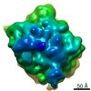
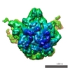
| Images | 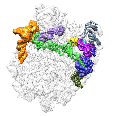 emd_0372.png emd_0372.png | 161.5 KB | ||
| Filedesc metadata | emd-0372.cif.gz | 14.2 KB | ||
| Archive directory |  http://ftp.pdbj.org/pub/emdb/structures/EMD-0372ftp://ftp.pdbj.org/pub/emdb/structures/EMD-0372 http://ftp.pdbj.org/pub/emdb/structures/EMD-0372ftp://ftp.pdbj.org/pub/emdb/structures/EMD-0372 | HTTPS FTP |
-Related structure data
| Related structure data |  6n8mMC 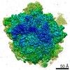 0369C 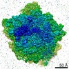 0370C 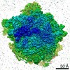 0371C 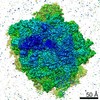 0373C 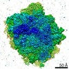 0374C 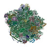 6n8jC 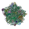 6n8kC 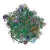 6n8lC 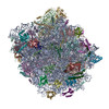 6n8nC 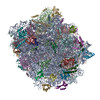 6n8oC C: citing same article ( M: atomic model generated by this map |
|---|---|
| Similar structure data |
-Links
| EMDB pages | EMDB (EBI/PDBe) / EMDataResource |
|---|---|
| Related items in Molecule of the Month |


-Map
| File | Download / File: emd_0372.map.gz / Format: CCP4 / Size: 216 MB / Type: IMAGE STORED AS FLOATING POINT NUMBER (4 BYTES) | ||||||||||||||||||||||||||||||||||||||||||||||||||||||||||||
|---|---|---|---|---|---|---|---|---|---|---|---|---|---|---|---|---|---|---|---|---|---|---|---|---|---|---|---|---|---|---|---|---|---|---|---|---|---|---|---|---|---|---|---|---|---|---|---|---|---|---|---|---|---|---|---|---|---|---|---|---|---|
| Annotation | Cryo-EM structure of pre-Lsg1 (PL) pre-60S ribosomal subunit | ||||||||||||||||||||||||||||||||||||||||||||||||||||||||||||
| Projections & slices | Image control
Images are generated by Spider. | ||||||||||||||||||||||||||||||||||||||||||||||||||||||||||||
| Voxel size | X=Y=Z: 1.1 Å | ||||||||||||||||||||||||||||||||||||||||||||||||||||||||||||
| Density |
| ||||||||||||||||||||||||||||||||||||||||||||||||||||||||||||
| Symmetry | Space group: 1 | ||||||||||||||||||||||||||||||||||||||||||||||||||||||||||||
| Details | EMDB XML:
CCP4 map header:
| ||||||||||||||||||||||||||||||||||||||||||||||||||||||||||||
 Z (Sec.)
 Y (Row.)
 X (Col.)


-Supplemental data
- Sample components
+Entire : Pre-Lsg1 (PL) pre-60S ribosomal subunit
+Supramolecule #1: Pre-Lsg1 (PL) pre-60S ribosomal subunit
+Macromolecule #1: Saccharomyces cerevisiae S288C 35S pre-ribosomal RNA (RDN37-1), m...
+Macromolecule #2: 5S rRNA
+Macromolecule #3: 5.8S RNA
+Macromolecule #4: Tyrosine-protein phosphatase YVH1
+Macromolecule #5: 60S ribosomal protein L2-A
+Macromolecule #6: 60S ribosomal protein L3
+Macromolecule #7: 60S ribosomal protein L4-A
+Macromolecule #8: 60S ribosomal protein L5
+Macromolecule #9: 60S ribosomal protein L6-A
+Macromolecule #10: 60S ribosomal protein L7-A
+Macromolecule #11: 60S ribosomal protein L8-A
+Macromolecule #12: 60S ribosomal protein L9-A
+Macromolecule #13: 60S ribosomal protein L11-A
+Macromolecule #14: 60S ribosomal protein L13-A
+Macromolecule #15: 60S ribosomal protein L14-A
+Macromolecule #16: 60S ribosomal protein L42-A
+Macromolecule #17: 60S ribosomal protein L43-A
+Macromolecule #18: Ribosomal Protein uL1
+Macromolecule #19: 60S ribosomal protein L15-A
+Macromolecule #20: 60S ribosomal protein L16-A
+Macromolecule #21: 60S ribosomal protein L17-A
+Macromolecule #22: 60S ribosomal protein L18-A
+Macromolecule #23: 60S ribosomal protein L19-A
+Macromolecule #24: 60S ribosomal protein L20-A
+Macromolecule #25: 60S ribosomal protein L21-A
+Macromolecule #26: 60S ribosomal protein L22-A
+Macromolecule #27: 60S ribosomal protein L23-A
+Macromolecule #28: 60S ribosomal protein L24-A
+Macromolecule #29: 60S ribosomal protein L25
+Macromolecule #30: 60S ribosomal protein L26-A
+Macromolecule #31: 60S ribosomal protein L27-A
+Macromolecule #32: 60S ribosomal protein L28
+Macromolecule #33: 60S ribosomal protein L29
+Macromolecule #34: 60S ribosomal protein L30
+Macromolecule #35: 60S ribosomal protein L31-A
+Macromolecule #36: 60S ribosomal protein L32
+Macromolecule #37: 60S ribosomal protein L33-A
+Macromolecule #38: 60S ribosomal protein L34-A
+Macromolecule #39: 60S ribosomal protein L35-A
+Macromolecule #40: 60S ribosomal protein L36-A
+Macromolecule #41: 60S ribosomal protein L37-A
+Macromolecule #42: 60S ribosomal protein L38
+Macromolecule #43: 60S ribosomal protein L39
+Macromolecule #44: Ubiquitin-60S ribosomal protein L40
+Macromolecule #45: 60S ribosomal export protein NMD3
+Macromolecule #46: Ribosomal protein L12
+Macromolecule #47: Eukaryotic translation initiation factor 6
-Experimental details
-Structure determination
| Method | cryo EM |
|---|---|
 Processing Processing | single particle reconstruction |
| Aggregation state | particle |
-Sample preparation
| Buffer | pH: 7.5 |
|---|---|
| Grid | Support film - Material: CARBON / Support film - topology: HOLEY / Details: unspecified |
| Vitrification | Cryogen name: ETHANE / Chamber humidity: 100 % / Instrument: FEI VITROBOT MARK IV |
- Electron microscopy
| Microscope | FEI TITAN KRIOS |
|---|---|
| Image recording | Film or detector model: GATAN K2 SUMMIT (4k x 4k) / Detector mode: COUNTING / Average exposure time: 6.0 sec. / Average electron dose: 40.0 e/Å2 |
| Electron beam | Acceleration voltage: 300 kV / Electron source:  FIELD EMISSION GUN FIELD EMISSION GUN |
| Electron optics | C2 aperture diameter: 100.0 µm / Illumination mode: FLOOD BEAM / Imaging mode: BRIGHT FIELD / Nominal defocus max: 2.5 µm / Nominal defocus min: 1.0 µm / Nominal magnification: 22500 |
| Sample stage | Specimen holder model: FEI TITAN KRIOS AUTOGRID HOLDER / Cooling holder cryogen: NITROGEN |
| Experimental equipment |  Model: Titan Krios / Image courtesy: FEI Company |