 ムービー
ムービー コントローラー


+ データを開く

- 基本情報
基本情報
| 登録情報 | 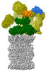 | ||||||||||||
|---|---|---|---|---|---|---|---|---|---|---|---|---|---|
| タイトル | Spinach 26S proteasome | ||||||||||||
 マップデータ マップデータ | |||||||||||||
 試料 試料 |
| ||||||||||||
 キーワード キーワード | PROTEASOME / UPS / PLANT / SPINACH / PLANT PROTEIN | ||||||||||||
| 機能・相同性 |  機能・相同性情報 機能・相同性情報nuclear proteasome complex / : / : / proteasome regulatory particle / cytosolic proteasome complex / proteasome regulatory particle, lid subcomplex / proteasome-activating activity / proteasome regulatory particle, base subcomplex / metal-dependent deubiquitinase activity / protein deubiquitination ...nuclear proteasome complex / : / : / proteasome regulatory particle / cytosolic proteasome complex / proteasome regulatory particle, lid subcomplex / proteasome-activating activity / proteasome regulatory particle, base subcomplex / metal-dependent deubiquitinase activity / protein deubiquitination / proteasome core complex / K63-linked deubiquitinase activity / proteasome binding / regulation of protein catabolic process / proteasome storage granule / polyubiquitin modification-dependent protein binding / proteasome endopeptidase complex / positive regulation of RNA polymerase II transcription preinitiation complex assembly / proteasome core complex, beta-subunit complex / proteasome core complex, alpha-subunit complex / proteasomal protein catabolic process / threonine-type endopeptidase activity / enzyme regulator activity / proteasome complex / proteolysis involved in protein catabolic process / protein catabolic process / metallopeptidase activity / peptidase activity / ubiquitin-dependent protein catabolic process / proteasome-mediated ubiquitin-dependent protein catabolic process / membrane => GO:0016020 / structural molecule activity / ATP hydrolysis activity / ATP binding / nucleus / cytoplasm / cytosol 類似検索 - 分子機能 | ||||||||||||
| 生物種 |  Spinacia oleracea (ホウレンソウ) Spinacia oleracea (ホウレンソウ) | ||||||||||||
| 手法 | 単粒子再構成法 / クライオ電子顕微鏡法 / 解像度: 3.3 Å | ||||||||||||
 データ登録者 データ登録者 | Kandolf S / Grishkovskaya I | ||||||||||||
| 資金援助 | 1件
| ||||||||||||
 引用 引用 | ジャーナル: Plant Commun / 年: 2022 タイトル: Cryo-EM structure of the plant 26S proteasome. 著者: Susanne Kandolf / Irina Grishkovskaya / Katarina Belačić / Derek L Bolhuis / Sascha Amann / Brent Foster / Richard Imre / Karl Mechtler / Alexander Schleiffer / Hemant D Tagare / Ellen D ...著者: Susanne Kandolf / Irina Grishkovskaya / Katarina Belačić / Derek L Bolhuis / Sascha Amann / Brent Foster / Richard Imre / Karl Mechtler / Alexander Schleiffer / Hemant D Tagare / Ellen D Zhong / Anton Meinhart / Nicholas G Brown / David Haselbach /    要旨: Targeted proteolysis is a hallmark of life. It is especially important in long-lived cells that can be found in higher eukaryotes, like plants. This task is mainly fulfilled by the ubiquitin- ...Targeted proteolysis is a hallmark of life. It is especially important in long-lived cells that can be found in higher eukaryotes, like plants. This task is mainly fulfilled by the ubiquitin-proteasome system. Thus, proteolysis by the 26S proteasome is vital to development, immunity, and cell division. Although the yeast and animal proteasomes are well characterized, there is only limited information on the plant proteasome. We determined the first plant 26S proteasome structure from Spinacia oleracea by single-particle electron cryogenic microscopy at an overall resolution of 3.3 Å. We found an almost identical overall architecture of the spinach proteasome compared with the known structures from mammals and yeast. Nevertheless, we noticed a structural difference in the proteolytic active β1 subunit. Furthermore, we uncovered an unseen compression state by characterizing the proteasome's conformational landscape. We suspect that this new conformation of the 20S core protease, in correlation with a partial opening of the unoccupied gate, may contribute to peptide release after proteolysis. Our data provide a structural basis for the plant proteasome, which is crucial for further studies. | ||||||||||||
| 履歴 |
|
- 構造の表示
構造の表示
| 添付画像 |
|---|

- ダウンロードとリンク
-EMDBアーカイブ
| マップデータ | emd_14175.map.gz | 22.4 MB | EMDBマップデータ形式 | |
|---|---|---|---|---|
| ヘッダ (付随情報) | emd-14175-v30.xmlemd-14175.xml | 30.1 KB 30.1 KB | 表示 表示 | EMDBヘッダ |
| 画像 | 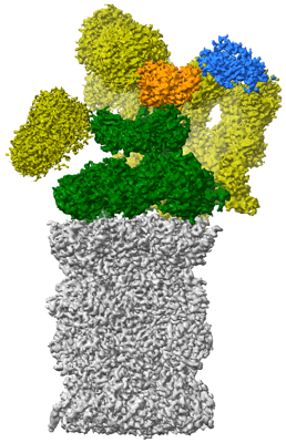 emd_14175.png emd_14175.png | 130.9 KB | ||
| Filedesc metadata | emd-14175.cif.gz | 8.5 KB | ||
| アーカイブディレクトリ |  http://ftp.pdbj.org/pub/emdb/structures/EMD-14175ftp://ftp.pdbj.org/pub/emdb/structures/EMD-14175 http://ftp.pdbj.org/pub/emdb/structures/EMD-14175ftp://ftp.pdbj.org/pub/emdb/structures/EMD-14175 | HTTPS FTP |
-関連構造データ
| 関連構造データ |  7qveMC  8amzM  7qvg M: このマップから作成された原子モデル C: 同じ文献を引用 ( |
|---|---|
| 類似構造データ | |
| 電子顕微鏡画像生データ | EMPIAR-10974 (タイトル: Cryo-EM structure of the plant 26S proteasome / Data size: 7.9 TB Data #1: Unaligned multi-frame micrographs of spinach 26S proteasome [micrographs - multiframe] Data #2: Unaligned multi-frame micrographs of spinach 26S proteasome [micrographs - multiframe] Data #3: Unaligned multi-frame micrographs of spinach 26S proteasome [micrographs - multiframe]) |
-リンク
| EMDBのページ | EMDB (EBI/PDBe) / EMDataResource |
|---|---|
| 「今月の分子」の関連する項目 |


-マップ
| ファイル | ダウンロード / ファイル: emd_14175.map.gz / 形式: CCP4 / 大きさ: 325 MB / タイプ: IMAGE STORED AS FLOATING POINT NUMBER (4 BYTES) | ||||||||||||||||||||
|---|---|---|---|---|---|---|---|---|---|---|---|---|---|---|---|---|---|---|---|---|---|
| ボクセルのサイズ | X=Y=Z: 1.23113 Å | ||||||||||||||||||||
| 密度 |
| ||||||||||||||||||||
| 対称性 | 空間群: 1 | ||||||||||||||||||||
| 詳細 | EMDB XML:
|
-添付データ
- 試料の構成要素
+全体 : Spinach 26S proteasome
+超分子 #1: Spinach 26S proteasome
+分子 #1: Proteasome subunit alpha type
+分子 #2: Proteasome subunit alpha type
+分子 #3: Proteasome subunit alpha type
+分子 #4: Proteasome subunit alpha type
+分子 #5: Proteasome subunit alpha type
+分子 #6: Proteasome subunit alpha type-3
+分子 #7: Proteasome subunit alpha type
+分子 #8: Proteasome subunit beta
+分子 #9: Proteasome subunit beta
+分子 #10: Proteasome subunit beta
+分子 #11: Proteasome subunit beta
+分子 #12: Proteasome subunit beta type-5
+分子 #13: Proteasome subunit beta
+分子 #14: Proteasome subunit beta
-実験情報
-構造解析
| 手法 | クライオ電子顕微鏡法 |
|---|---|
 解析 解析 | 単粒子再構成法 |
| 試料の集合状態 | particle |
-試料調製
| 濃度 | 0.02 mg/mL | |||||||||||||||||||||||||||
|---|---|---|---|---|---|---|---|---|---|---|---|---|---|---|---|---|---|---|---|---|---|---|---|---|---|---|---|---|
| 緩衝液 | pH: 6.5 構成要素:
| |||||||||||||||||||||||||||
| グリッド | モデル: Quantifoil R3.5/1 / 材質: COPPER / メッシュ: 200 / 支持フィルム - 材質: CARBON / 支持フィルム - トポロジー: CONTINUOUS / 支持フィルム - Film thickness: 2 / 前処理 - タイプ: GLOW DISCHARGE / 前処理 - 時間: 60 sec. / 前処理 - 雰囲気: AIR | |||||||||||||||||||||||||||
| 凍結 | 凍結剤: ETHANE / チャンバー内湿度: 80 % / チャンバー内温度: 277 K / 装置: LEICA EM GP |
- 電子顕微鏡法
| 顕微鏡 | FEI TITAN KRIOS |
|---|---|
| 撮影 | #0 - Image recording ID: 1 #0 - フィルム・検出器のモデル: FEI FALCON III (4k x 4k) #0 - 検出モード: INTEGRATING / #0 - 実像数: 24769 / #0 - 平均電子線量: 80.0 e/Å2 / #1 - Image recording ID: 2 #1 - フィルム・検出器のモデル: FEI FALCON III (4k x 4k) #1 - 検出モード: INTEGRATING / #1 - デジタル化 - サイズ - 横: 4096 pixel / #1 - デジタル化 - サイズ - 縦: 4096 pixel / #1 - 実像数: 8089 / #1 - 平均電子線量: 50.0 e/Å2 |
| 電子線 | 加速電圧: 300 kV / 電子線源:  FIELD EMISSION GUN FIELD EMISSION GUN |
| 電子光学系 | C2レンズ絞り径: 70.0 µm / 照射モード: FLOOD BEAM / 撮影モード: BRIGHT FIELD / Cs: 2.7 mm / 最大 デフォーカス(公称値): 4.0 µm / 最小 デフォーカス(公称値): 1.0 µm |
| 試料ステージ | 試料ホルダーモデル: FEI TITAN KRIOS AUTOGRID HOLDER ホルダー冷却材: NITROGEN |
| 実験機器 |  モデル: Titan Krios / 画像提供: FEI Company |

