Translation initiation factor 3 / Translation initiation factor 3, N-terminal / Translation initiation factor 3 (IF-3), N-terminal domain superfamily / Translation initiation factor 3 (IF-3), C-terminal domain superfamily / Translation initiation factor IF-3, N-terminal domain / Coiled-coil-helix-coiled-coil-helix domain-containing protein 1 / 28S ribosomal protein S26 / Mitochondrial ribosome subunit S26 / Cysteine alpha-hairpin motif superfamily / PPR repeat family ...Translation initiation factor 3 / Translation initiation factor 3, N-terminal / Translation initiation factor 3 (IF-3), N-terminal domain superfamily / Translation initiation factor 3 (IF-3), C-terminal domain superfamily / Translation initiation factor IF-3, N-terminal domain / Coiled-coil-helix-coiled-coil-helix domain-containing protein 1 / 28S ribosomal protein S26 / Mitochondrial ribosome subunit S26 / Cysteine alpha-hairpin motif superfamily / PPR repeat family / 28S ribosomal protein S27, mitochondrial / MRPS27/PTCD2 / : / Mitochondrial 28S ribosomal protein S27 / Ribosomal protein S22, mitochondrial / Ribosomal protein S23, mitochondrial / Ribosomal protein S23/S25, mitochondrial / Mitochondrial 28S ribosomal protein S31 / Mitochondrial 28S ribosomal protein S22 / Mitochondrial ribosomal protein S23 / Mitochondrial 28S ribosomal protein S31 / Ribosomal protein S29, mitochondrial / Ribosomal protein S28, mitochondrial / 28S ribosomal protein S10, mitochondrial / Mitochondrial ribosomal protein MRP-S35 / 28S ribosomal protein S24, mitochondrial / Mitochondrial 28S ribosomal protein S34 / Pentatricopeptide repeat domain-containing protein 3 / 28S ribosomal protein S17, mitochondrial / 28S ribosomal protein S18b, mitochondrial / : / Mitochondrial ribosome subunit S24 / Mitochondrial 28S ribosomal protein S34 / Small ribosomal subunit protein uS5m, N-terminal / 28S ribosomal protein S25, mitochondrial / Pentatricopeptide repeat domain / Ribosomal protein S27/S33, mitochondrial / Ribosomal protein S24/S35, mitochondrial / Mitochondrial ribosomal subunit S27 / Ribosomal protein S24/S35, mitochondrial, conserved domain / Mitochondrial ribosomal subunit protein / Pentatricopeptide (PPR) repeat profile. / Ribosomal protein S23/S29, mitochondrial / Mitochondrial ribosomal death-associated protein 3 / Pentatricopeptide repeat / Mitochondrial mRNA-processing protein COX24, C-terminal / Mitochondrial mRNA-processing protein COX24, C-terminal / Mitochondrial domain of unknown function (DUF1713) / CHCH / CHCH domain / Coiled coil-helix-coiled coil-helix (CHCH) domain profile. / Ribosomal protein/NADH dehydrogenase domain / Mitochondrial ribosomal protein L51 / S25 / CI-B8 domain / Mitochondrial ribosomal protein L51 / S25 / CI-B8 domain / Ribosomal protein S21 superfamily / Ribosomal protein S21 / Ribosomal protein S21 / Ribosomal protein S12, bacterial-type / Ribosomal protein S2, bacteria/mitochondria/plastid / Ribosomal protein S18, conserved site / Ribosomal protein S18 signature. / Ribosomal protein S16 / Ribosomal protein S16 / Ribosomal protein S16 domain superfamily / Ribosomal protein S15, bacterial-type / Ribosomal protein S6 / Ribosomal protein S6 / Ribosomal protein S6 superfamily / Translation elongation factor EF1B/ribosomal protein S6 / Ribosomal protein S18 / Ribosomal protein S18 / Ribosomal protein S18 superfamily / Ribosomal protein S12 signature. / Ribosomal protein S2 signature 1. / Ribosomal protein S12/S23 / Ribosomal protein S5, N-terminal, conserved site / Ribosomal protein S5 signature. / Ribosomal protein S2, conserved site / Ribosomal protein S2 / Ribosomal protein S2, flavodoxin-like domain superfamily / Ribosomal protein S2 / S5 double stranded RNA-binding domain profile. / Ribosomal protein S5 / Ribosomal protein S5, N-terminal / Ribosomal protein S5, N-terminal domain / Ribosomal protein S5, C-terminal / Ribosomal protein S5, C-terminal domain / Ribosomal protein S14 / Ribosomal protein S14p/S29e / Ribosomal protein S12/S23 / Ribosomal S11, conserved site / Ribosomal protein S11 signature. / Ribosomal protein S10p/S20e / Ribosomal protein S11 / Ribosomal protein S10 domain / Ribosomal protein S10 domain superfamily / Ribosomal protein S10p/S20e / Ribosomal protein S9, conserved site / Ribosomal protein S9 signature. / Ribosomal protein S11 Similarity search - Domain/homology
Small ribosomal subunit protein uS12m / Small ribosomal subunit protein uS14m / Small ribosomal subunit protein mS29 / Small ribosomal subunit protein mS22 / Small ribosomal subunit protein mS25 / Small ribosomal subunit protein uS10m / Small ribosomal subunit protein mS35 / Small ribosomal subunit protein uS5m / Small ribosomal subunit protein uS11m / Small ribosomal subunit protein uS15m ...Small ribosomal subunit protein uS12m / Small ribosomal subunit protein uS14m / Small ribosomal subunit protein mS29 / Small ribosomal subunit protein mS22 / Small ribosomal subunit protein mS25 / Small ribosomal subunit protein uS10m / Small ribosomal subunit protein mS35 / Small ribosomal subunit protein uS5m / Small ribosomal subunit protein uS11m / Small ribosomal subunit protein uS15m / Small ribosomal subunit protein bS21m / Small ribosomal subunit protein mS34 / Small ribosomal subunit protein bS6m / Small ribosomal subunit protein uS9m / Small ribosomal subunit protein mS27 / Small ribosomal subunit protein mS31 / Small ribosomal subunit protein mS37 / Small ribosomal subunit protein uS3m / Small ribosomal subunit protein mS39 / Small ribosomal subunit protein mS26 / Translation initiation factor IF-3, mitochondrial / Small ribosomal subunit protein mS38 / Small ribosomal subunit protein mS33 / Small ribosomal subunit protein bS1m / Small ribosomal subunit protein uS17m / Small ribosomal subunit protein uS7m / Small ribosomal subunit protein uS2m / Small ribosomal subunit protein bS16m / Small ribosomal subunit protein bS18m / Small ribosomal subunit protein mS23 / Small ribosomal subunit protein mS40 Similarity search - Component
Biological species
Homo sapiens (human)
Method
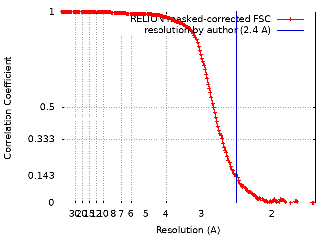
single particle reconstruction / cryo EM / Resolution: 2.4 Å
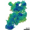
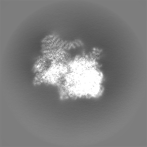
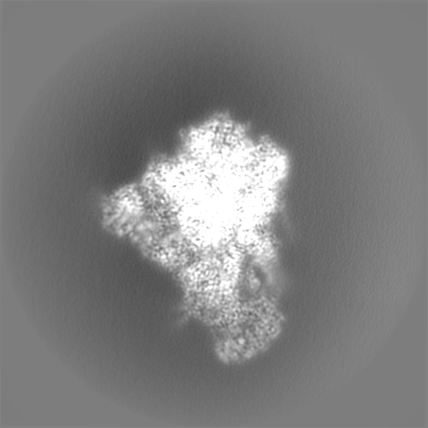
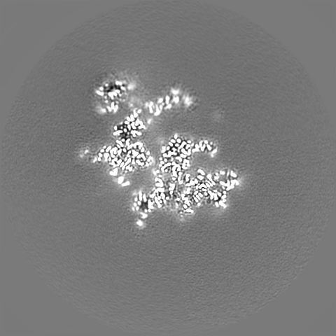
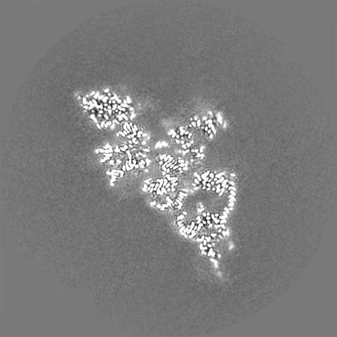
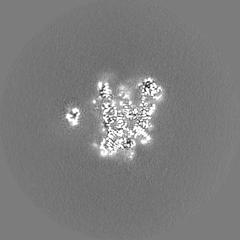
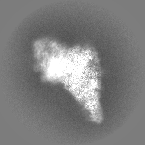
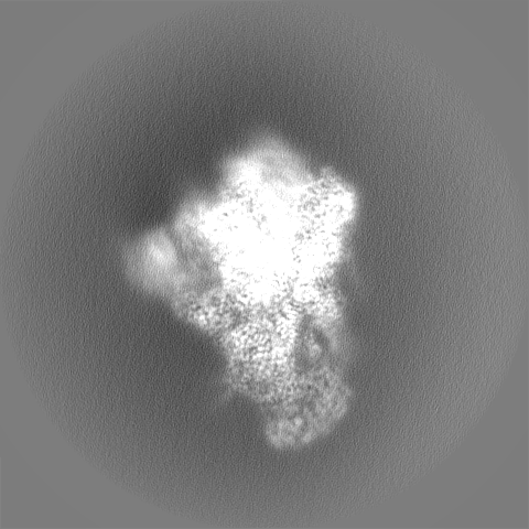
Journal: Elife / Year: 2022 Title: Structure of the mitoribosomal small subunit with streptomycin reveals Fe-S clusters and physiological molecules. Authors: Yuzuru Itoh / Vivek Singh / Anas Khawaja / Andreas Naschberger / Minh Duc Nguyen / Joanna Rorbach / Alexey Amunts / Abstract: The mitoribosome regulates cellular energy production, and its dysfunction is associated with aging. Inhibition of the mitoribosome can be caused by off-target binding of antimicrobial drugs and was ...The mitoribosome regulates cellular energy production, and its dysfunction is associated with aging. Inhibition of the mitoribosome can be caused by off-target binding of antimicrobial drugs and was shown to be coupled with a bilateral decreased visual acuity. Previously, we reported mitochondria-specific protein aspects of the mitoribosome, and in this article we present a 2.4-Å resolution structure of the small subunit in a complex with the anti-tuberculosis drug streptomycin that reveals roles of non-protein components. We found iron-sulfur clusters that are coordinated by different mitoribosomal proteins, nicotinamide adenine dinucleotide (NAD) associated with rRNA insertion, and posttranslational modifications. This is the first evidence of inter-protein coordination of iron-sulfur, and the finding of iron-sulfur clusters and NAD as fundamental building blocks of the mitoribosome directly links to mitochondrial disease and aging. We also report details of streptomycin interactions, suggesting that the mitoribosome-bound streptomycin is likely to be in hydrated gem-diol form and can be subjected to other modifications by the cellular milieu. The presented approach of adding antibiotics to cultured cells can be used to define their native structures in a bound form under more physiological conditions, and since streptomycin is a widely used drug for treatment, the newly resolved features can serve as determinants for targeting.
Model: Quantifoil R2/1 / Material: COPPER / Support film - Material: CARBON / Support film - topology: CONTINUOUS / Support film - Film thickness: 3 / Pretreatment - Type: GLOW DISCHARGE / Pretreatment - Time: 30 sec.
Vitrification
Cryogen name: ETHANE / Chamber humidity: 100 % / Chamber temperature: 297 K / Instrument: FEI VITROBOT MARK IV
-
Electron microscopy
Microscope
FEI TITAN KRIOS
Specialist optics
Energy filter - Name: GIF Quantum LS / Energy filter - Slit width: 20 eV
Image recording
Film or detector model: GATAN K2 SUMMIT (4k x 4k) / Detector mode: COUNTING / Digitization - Frames/image: 1-20 / Number real images: 31773 / Average exposure time: 4.0 sec. / Average electron dose: 30.0 e/Å2
Electron beam
Acceleration voltage: 300 kV / Electron source: FIELD EMISSION GUN
In the structure databanks used in Yorodumi, some data are registered as the other names, "COVID-19 virus" and "2019-nCoV". Here are the details of the virus and the list of structure data.
Jan 31, 2019. EMDB accession codes are about to change! (news from PDBe EMDB page)
EMDB accession codes are about to change! (news from PDBe EMDB page)
The allocation of 4 digits for EMDB accession codes will soon come to an end. Whilst these codes will remain in use, new EMDB accession codes will include an additional digit and will expand incrementally as the available range of codes is exhausted. The current 4-digit format prefixed with “EMD-” (i.e. EMD-XXXX) will advance to a 5-digit format (i.e. EMD-XXXXX), and so on. It is currently estimated that the 4-digit codes will be depleted around Spring 2019, at which point the 5-digit format will come into force.
The EM Navigator/Yorodumi systems omit the EMD- prefix.
Related info.:Q: What is EMD? / ID/Accession-code notation in Yorodumi/EM Navigator
Yorodumi is a browser for structure data from EMDB, PDB, SASBDB, etc.
This page is also the successor to EM Navigator detail page, and also detail information page/front-end page for Omokage search.
The word "yorodu" (or yorozu) is an old Japanese word meaning "ten thousand". "mi" (miru) is to see.
Related info.:EMDB / PDB / SASBDB / Comparison of 3 databanks / Yorodumi Search / Aug 31, 2016. New EM Navigator & Yorodumi / Yorodumi Papers / Jmol/JSmol / Function and homology information / Changes in new EM Navigator and Yorodumi
 Movie
Movie Controller

 Yorodumi
 Open data
 Basic information
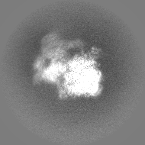
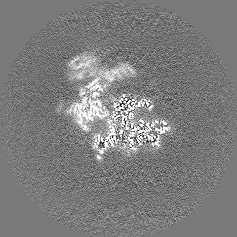
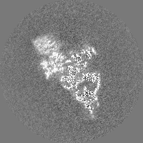
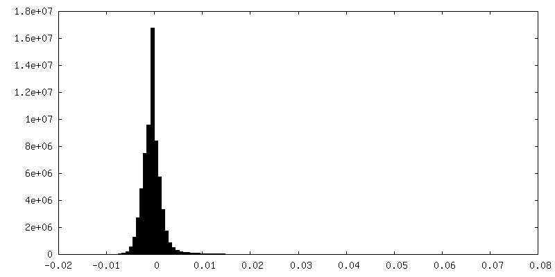
 Map data
 Sample
 Keywords
 Function and homology information
 Homo sapiens (human)
 Authors
 Sweden, 2 items
  Citation
 Structure visualization
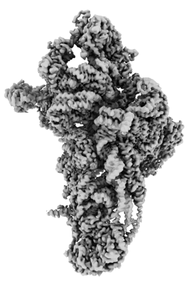
 Downloads & links
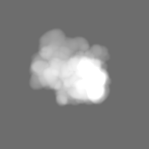
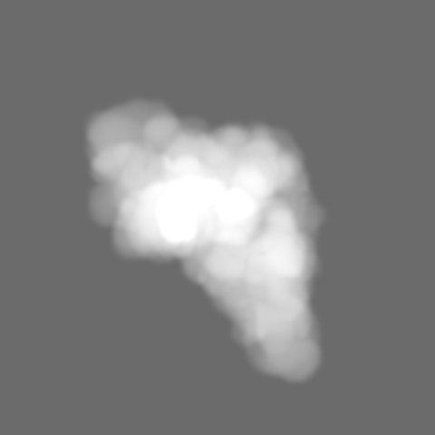
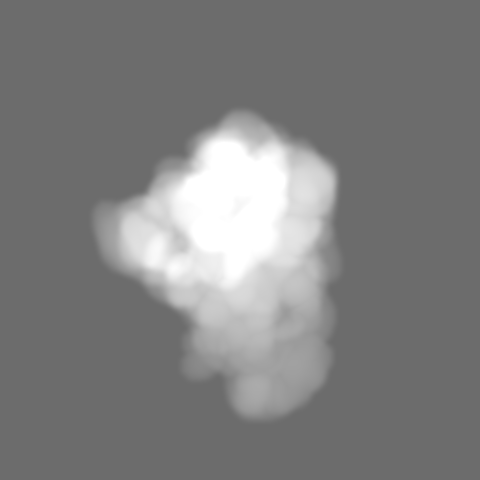
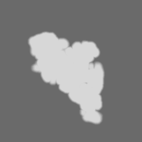
 emd_13170.png
 http://ftp.pdbj.org/pub/emdb/structures/EMD-13170


 Z
Z Y
Y X
X


 Sample components


 Processing
 Electron microscopy
 FIELD EMISSION GUN