 Movie
Movie Controller


+ Open data

- Basic information
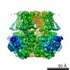
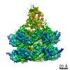
| Entry | Database: EMDB / ID: EMD-12448 | |||||||||
|---|---|---|---|---|---|---|---|---|---|---|
| Title | diazaborine bound Drg1(AFG2) | |||||||||
 Map data Map data | ||||||||||
 Sample Sample |
| |||||||||
 Keywords Keywords | DRG1 / AFG2 / ribosome maturation / AAA protein / diazaborine / inhibitor / RIBOSOME | |||||||||
| Function / homology |  Function and homology information Function and homology informationprotein hexamerization / non-chaperonin molecular chaperone ATPase / preribosome, large subunit precursor / ribosomal large subunit biogenesis / response to xenobiotic stimulus / ATP hydrolysis activity / ATP binding / cytoplasm Similarity search - Function | |||||||||
| Biological species |  | |||||||||
| Method | single particle reconstruction / cryo EM / Resolution: 3.4 Å | |||||||||
 Authors Authors | Prattes M / Hodirnau VV / Grishkovskaya I / Bergler H / Haselbach D | |||||||||
 Citation Citation | Journal: Nat Commun / Year: 2021 Title: Structural basis for inhibition of the AAA-ATPase Drg1 by diazaborine. Authors: Michael Prattes / Irina Grishkovskaya / Victor-Valentin Hodirnau / Ingrid Rössler / Isabella Klein / Christina Hetzmannseder / Gertrude Zisser / Christian C Gruber / Karl Gruber / David ...Authors: Michael Prattes / Irina Grishkovskaya / Victor-Valentin Hodirnau / Ingrid Rössler / Isabella Klein / Christina Hetzmannseder / Gertrude Zisser / Christian C Gruber / Karl Gruber / David Haselbach / Helmut Bergler /  Abstract: The hexameric AAA-ATPase Drg1 is a key factor in eukaryotic ribosome biogenesis and initiates cytoplasmic maturation of the large ribosomal subunit by releasing the shuttling maturation factor Rlp24. ...The hexameric AAA-ATPase Drg1 is a key factor in eukaryotic ribosome biogenesis and initiates cytoplasmic maturation of the large ribosomal subunit by releasing the shuttling maturation factor Rlp24. Drg1 monomers contain two AAA-domains (D1 and D2) that act in a concerted manner. Rlp24 release is inhibited by the drug diazaborine which blocks ATP hydrolysis in D2. The mode of inhibition was unknown. Here we show the first cryo-EM structure of Drg1 revealing the inhibitory mechanism. Diazaborine forms a covalent bond to the 2'-OH of the nucleotide in D2, explaining its specificity for this site. As a consequence, the D2 domain is locked in a rigid, inactive state, stalling the whole Drg1 hexamer. Resistance mechanisms identified include abolished drug binding and altered positioning of the nucleotide. Our results suggest nucleotide-modifying compounds as potential novel inhibitors for AAA-ATPases. | |||||||||
| History |
|
- Structure visualization
| Movie |
Movie viewer |
|---|---|
| Structure viewer | EM map: SurfViewMolmilJmol/JSmol |
| Supplemental images |

- Downloads & links
-EMDB archive
| Map data | emd_12448.map.gz | 48.5 MB | EMDB map data format | |
|---|---|---|---|---|
| Header (meta data) | emd-12448-v30.xmlemd-12448.xml | 23.2 KB 23.2 KB | Display Display | EMDB header |
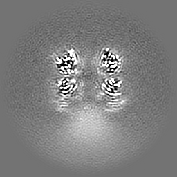
| Images |  emd_12448.png emd_12448.png | 211.7 KB | ||
| Filedesc metadata | emd-12448.cif.gz | 7.1 KB | ||
| Others | emd_12448_additional_1.map.gzemd_12448_additional_2.map.gz | 57.2 MB 57.8 MB | ||
| Archive directory |  http://ftp.pdbj.org/pub/emdb/structures/EMD-12448ftp://ftp.pdbj.org/pub/emdb/structures/EMD-12448 http://ftp.pdbj.org/pub/emdb/structures/EMD-12448ftp://ftp.pdbj.org/pub/emdb/structures/EMD-12448 | HTTPS FTP |
-Related structure data
| Related structure data |  7nkuMC M: atomic model generated by this map C: citing same article ( |
|---|---|
| Similar structure data | |
| EM raw data | EMPIAR-10717 (Title: Single particle Data of diazaborine bound Drg1 / Data size: 1.6 TB Data #1: Unaligned Multiframe micrographs of DRG1 bound to diazaborine. [micrographs - multiframe]) |


-Links
| EMDB pages | EMDB (EBI/PDBe) / EMDataResource |
|---|---|
| Related items in Molecule of the Month |


-Map
| File | Download / File: emd_12448.map.gz / Format: CCP4 / Size: 64 MB / Type: IMAGE STORED AS FLOATING POINT NUMBER (4 BYTES) | ||||||||||||||||||||||||||||||||||||||||||||||||||||||||||||
|---|---|---|---|---|---|---|---|---|---|---|---|---|---|---|---|---|---|---|---|---|---|---|---|---|---|---|---|---|---|---|---|---|---|---|---|---|---|---|---|---|---|---|---|---|---|---|---|---|---|---|---|---|---|---|---|---|---|---|---|---|---|
| Projections & slices | Image control
Images are generated by Spider. | ||||||||||||||||||||||||||||||||||||||||||||||||||||||||||||
| Voxel size | X=Y=Z: 1.06 Å | ||||||||||||||||||||||||||||||||||||||||||||||||||||||||||||
| Density |
| ||||||||||||||||||||||||||||||||||||||||||||||||||||||||||||
| Symmetry | Space group: 1 | ||||||||||||||||||||||||||||||||||||||||||||||||||||||||||||
| Details | EMDB XML:
CCP4 map header:
| ||||||||||||||||||||||||||||||||||||||||||||||||||||||||||||
 Z (Sec.)
 Y (Row.)
 X (Col.)


-Supplemental data
-Additional map: #2
| File | emd_12448_additional_1.map | ||||||||||||
|---|---|---|---|---|---|---|---|---|---|---|---|---|---|
| Projections & Slices |
| ||||||||||||
| Density Histograms |


-Additional map: #1
| File | emd_12448_additional_2.map | ||||||||||||
|---|---|---|---|---|---|---|---|---|---|---|---|---|---|
| Projections & Slices |
| ||||||||||||
| Density Histograms |


- Sample components
-Entire : Drg1 (AFG2) bound to diazaborine
| Entire | Name: Drg1 (AFG2) bound to diazaborine |
|---|---|
| Components |
|
-Supramolecule #1: Drg1 (AFG2) bound to diazaborine
| Supramolecule | Name: Drg1 (AFG2) bound to diazaborine / type: complex / ID: 1 / Parent: 0 / Macromolecule list: #1 |
|---|---|
| Source (natural) | Organism: |
-Macromolecule #1: ATPase family gene 2 protein
| Macromolecule | Name: ATPase family gene 2 protein / type: protein_or_peptide / ID: 1 / Number of copies: 6 / Enantiomer: LEVO / EC number: non-chaperonin molecular chaperone ATPase |
|---|---|
| Source (natural) | Organism: Strain: ATCC 204508 / S288c |
| Molecular weight | Theoretical: 84.850719 KDa |
| Recombinant expression | Organism:  |
| Sequence | String: MAPKSSSSGS KKKSSASSNS ADAKASKFKL PAEFITRPHP SKDHGKETCT AYIHPNVLSS LEINPGSFCT VGKIGENGIL VIARAGDEE VHPVNVITLS TTIRSVGNLI LGDRLELKKA QVQPPYATKV TVGSLQGYNI LECMEEKVIQ KLLDDSGVIM P GMIFQNLK ...String: MAPKSSSSGS KKKSSASSNS ADAKASKFKL PAEFITRPHP SKDHGKETCT AYIHPNVLSS LEINPGSFCT VGKIGENGIL VIARAGDEE VHPVNVITLS TTIRSVGNLI LGDRLELKKA QVQPPYATKV TVGSLQGYNI LECMEEKVIQ KLLDDSGVIM P GMIFQNLK TKAGDESIDV VITDASDDSL PDVSQLDLNM DDMYGGLDNL FYLSPPFIFR KGSTHITFSK ETQANRKYNL PE PLSYAAV GGLDKEIESL KSAIEIPLHQ PTLFSSFGVS PPRGILLHGP PGTGKTMLLR VVANTSNAHV LTINGPSIVS KYL GETEAA LRDIFNEARK YQPSIIFIDE IDSIAPNRAN DDSGEVESRV VATLLTLMDG MGAAGKVVVI AATNRPNSVD PALR RPGRF DQEVEIGIPD VDARFDILTK QFSRMSSDRH VLDSEAIKYI ASKTHGYVGA DLTALCRESV MKTIQRGLGT DANID KFSL KVTLKDVESA MVDIRPSAMR EIFLEMPKVY WSDIGGQEEL KTKMKEMIQL PLEASETFAR LGISAPKGVL LYGPPG CSK TLTAKALATE SGINFLAVKG PEIFNKYVGE SERAIREIFR KARSAAPSII FFDEIDALSP DRDGSSTSAA NHVLTSL LN EIDGVEELKG VVIVAATNRP DEIDAALLRP GRLDRHIYVG PPDVNARLEI LKKCTKKFNT EESGVDLHEL ADRTEGYS G AEVVLLCQEA GLAAIMEDLD VAKVELRHFE KAFKGIARGI TPEMLSYYEE FALRSGSSS UniProtKB: ATPase family gene 2 protein |
-Macromolecule #2: PHOSPHOTHIOPHOSPHORIC ACID-ADENYLATE ESTER
| Macromolecule | Name: PHOSPHOTHIOPHOSPHORIC ACID-ADENYLATE ESTER / type: ligand / ID: 2 / Number of copies: 12 / Formula: AGS |
|---|---|
| Molecular weight | Theoretical: 523.247 Da |
| Chemical component information |  ChemComp-AGS: |
-Macromolecule #3: 6-METHYL-2(PROPANE-1-SULFONYL)-2H-THIENO[3,2-D][1,2,3]DIAZABORINI...
| Macromolecule | Name: 6-METHYL-2(PROPANE-1-SULFONYL)-2H-THIENO[3,2-D][1,2,3]DIAZABORININ-1-OL type: ligand / ID: 3 / Number of copies: 6 / Formula: TDB |
|---|---|
| Molecular weight | Theoretical: 272.152 Da |
| Chemical component information |  ChemComp-TDB: |
-Experimental details
-Structure determination
| Method | cryo EM |
|---|---|
 Processing Processing | single particle reconstruction |
| Aggregation state | particle |
-Sample preparation
| Buffer | pH: 7.6 Component:
| |||||||||||||||||||||
|---|---|---|---|---|---|---|---|---|---|---|---|---|---|---|---|---|---|---|---|---|---|---|
| Grid | Model: Quantifoil R1.2/1.3 / Material: COPPER / Mesh: 200 / Support film - Material: CARBON / Support film - topology: HOLEY / Pretreatment - Type: GLOW DISCHARGE / Pretreatment - Time: 60 sec. | |||||||||||||||||||||
| Vitrification | Cryogen name: ETHANE |
- Electron microscopy
| Microscope | FEI TITAN KRIOS |
|---|---|
| Specialist optics | Energy filter - Name: GIF Bioquantum |
| Image recording | Film or detector model: GATAN K3 BIOQUANTUM (6k x 4k) / Average electron dose: 60.0 e/Å2 |
| Electron beam | Acceleration voltage: 300 kV / Electron source:  FIELD EMISSION GUN FIELD EMISSION GUN |
| Electron optics | Illumination mode: FLOOD BEAM / Imaging mode: BRIGHT FIELD |
| Sample stage | Specimen holder model: FEI TITAN KRIOS AUTOGRID HOLDER / Cooling holder cryogen: NITROGEN |
| Experimental equipment |  Model: Titan Krios / Image courtesy: FEI Company |
+Image processing
-Atomic model buiding 1
| Refinement | Space: REAL / Protocol: AB INITIO MODEL / Target criteria: correlation coefficient |
|---|---|
| Output model | PDB-7nku: |
