 Movie
Movie Controller


+ Open data

- Basic information
| Entry | Database: EMDB / ID: EMD-12353 | |||||||||
|---|---|---|---|---|---|---|---|---|---|---|
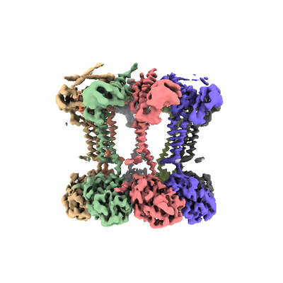
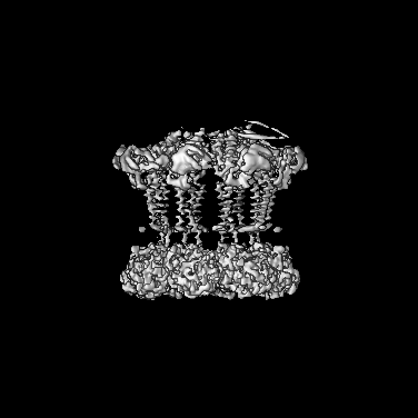
| Title | Wzc-K540M-4YE C1 | |||||||||
 Map data Map data | ||||||||||
 Sample Sample |
| |||||||||
 Keywords Keywords | Wzc / regulator / capsular polysaccharide synthesis and transport / Gram-negative pathogens / CARBOHYDRATE | |||||||||
| Function / homology | :  Function and homology information Function and homology information | |||||||||
| Biological species |  | |||||||||
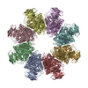
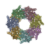
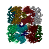
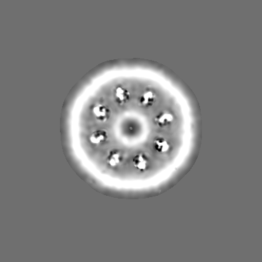
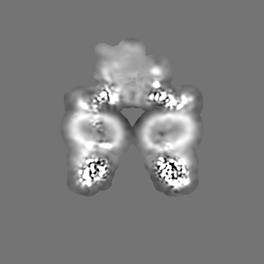
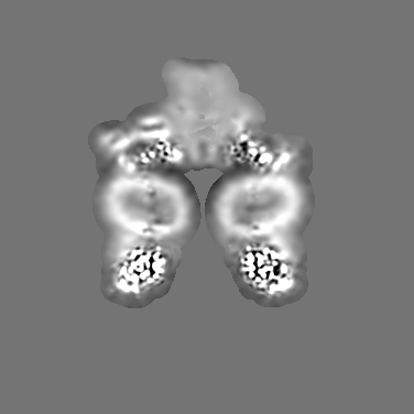
| Method | single particle reconstruction / cryo EM / Resolution: 3.51 Å | |||||||||
 Authors Authors | Naismith JH / Liu JW | |||||||||
| Funding support |  United Kingdom, 1 items United Kingdom, 1 items
| |||||||||
 Citation Citation | Journal: Nat Commun / Year: 2021 Title: The molecular basis of regulation of bacterial capsule assembly by Wzc. Authors: Yun Yang / Jiwei Liu / Bradley R Clarke / Laura Seidel / Jani R Bolla / Philip N Ward / Peijun Zhang / Carol V Robinson / Chris Whitfield / James H Naismith /  Abstract: Bacterial extracellular polysaccharides (EPSs) play critical roles in virulence. Many bacteria assemble EPSs via a multi-protein "Wzx-Wzy" system, involving glycan polymerization at the outer face of ...Bacterial extracellular polysaccharides (EPSs) play critical roles in virulence. Many bacteria assemble EPSs via a multi-protein "Wzx-Wzy" system, involving glycan polymerization at the outer face of the cytoplasmic/inner membrane. Gram-negative species couple polymerization with translocation across the periplasm and outer membrane and the master regulator of the system is the tyrosine autokinase, Wzc. This near atomic cryo-EM structure of dephosphorylated Wzc from E. coli shows an octameric assembly with a large central cavity formed by transmembrane helices. The tyrosine autokinase domain forms the cytoplasm region, while the periplasmic region contains small folded motifs and helical bundles. The helical bundles are essential for function, most likely through interaction with the outer membrane translocon, Wza. Autophosphorylation of the tyrosine-rich C-terminus of Wzc results in disassembly of the octamer into multiply phosphorylated monomers. We propose that the cycling between phosphorylated monomer and dephosphorylated octamer regulates glycan polymerization and translocation. | |||||||||
| History |
|
- Structure visualization
| Movie |
Movie viewer |
|---|---|
| Structure viewer | EM map: SurfViewMolmilJmol/JSmol |
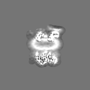
| Supplemental images |
- Downloads & links
-EMDB archive
| Map data | emd_12353.map.gz | 18 MB | EMDB map data format | |
|---|---|---|---|---|
| Header (meta data) | emd-12353-v30.xmlemd-12353.xml | 9.9 KB 9.9 KB | Display Display | EMDB header |
| Images |  emd_12353.png emd_12353.png | 94.6 KB | ||
| Filedesc metadata | emd-12353.cif.gz | 5.3 KB | ||
| Archive directory |  http://ftp.pdbj.org/pub/emdb/structures/EMD-12353ftp://ftp.pdbj.org/pub/emdb/structures/EMD-12353 http://ftp.pdbj.org/pub/emdb/structures/EMD-12353ftp://ftp.pdbj.org/pub/emdb/structures/EMD-12353 | HTTPS FTP |
-Validation report
| Summary document | emd_12353_validation.pdf.gz | 402 KB | Display | EMDB validaton report |
|---|---|---|---|---|
| Full document | emd_12353_full_validation.pdf.gz | 401.5 KB | Display | |
| Data in XML | emd_12353_validation.xml.gz | 7.2 KB | Display | |
| Data in CIF | emd_12353_validation.cif.gz | 8.2 KB | Display | |
| Arichive directory | https://ftp.pdbj.org/pub/emdb/validation_reports/EMD-12353ftp://ftp.pdbj.org/pub/emdb/validation_reports/EMD-12353 | HTTPS FTP |
-Related structure data
| Related structure data |  7nibMC  7nhrC  7nhsC  7ni2C  7nihC  7niiC M: atomic model generated by this map C: citing same article ( |
|---|---|
| Similar structure data |


-Links
| EMDB pages | EMDB (EBI/PDBe) / EMDataResource |
|---|
-Map
| File | Download / File: emd_12353.map.gz / Format: CCP4 / Size: 202.8 MB / Type: IMAGE STORED AS FLOATING POINT NUMBER (4 BYTES) | ||||||||||||||||||||||||||||||||||||||||||||||||||||||||||||||||||||
|---|---|---|---|---|---|---|---|---|---|---|---|---|---|---|---|---|---|---|---|---|---|---|---|---|---|---|---|---|---|---|---|---|---|---|---|---|---|---|---|---|---|---|---|---|---|---|---|---|---|---|---|---|---|---|---|---|---|---|---|---|---|---|---|---|---|---|---|---|---|
| Projections & slices | Image control
Images are generated by Spider. | ||||||||||||||||||||||||||||||||||||||||||||||||||||||||||||||||||||
| Voxel size | X=Y=Z: 0.831 Å | ||||||||||||||||||||||||||||||||||||||||||||||||||||||||||||||||||||
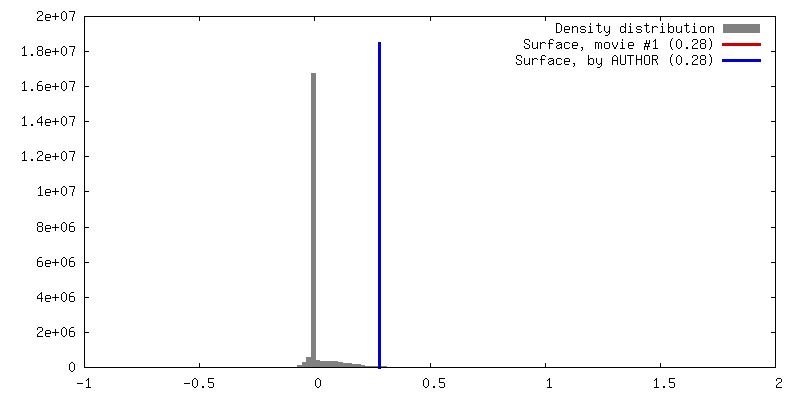
| Density |
| ||||||||||||||||||||||||||||||||||||||||||||||||||||||||||||||||||||
| Symmetry | Space group: 1 | ||||||||||||||||||||||||||||||||||||||||||||||||||||||||||||||||||||
| Details | EMDB XML:
CCP4 map header:
| ||||||||||||||||||||||||||||||||||||||||||||||||||||||||||||||||||||
 Z (Sec.)
 Y (Row.)
 X (Col.)


-Supplemental data
- Sample components
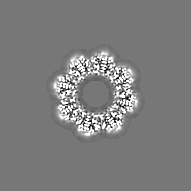
-Entire : Octameric complex of Wzc-K540M-4YE
| Entire | Name: Octameric complex of Wzc-K540M-4YE |
|---|---|
| Components |
|
-Supramolecule #1: Octameric complex of Wzc-K540M-4YE
| Supramolecule | Name: Octameric complex of Wzc-K540M-4YE / type: complex / ID: 1 / Parent: 0 / Macromolecule list: all |
|---|---|
| Source (natural) | Organism: |
| Molecular weight | Theoretical: 640 KDa |
-Macromolecule #1: Tyrosine-protein kinase
| Macromolecule | Name: Tyrosine-protein kinase / type: protein_or_peptide / ID: 1 / Number of copies: 8 / Enantiomer: LEVO / EC number: non-specific protein-tyrosine kinase |
|---|---|
| Source (natural) | Organism: |
| Molecular weight | Theoretical: 80.435109 KDa |
| Recombinant expression | Organism: |
| Sequence | String: MTSVTSKQST ILGSDEIDLG RVIGELIDHR KLIISITSVF TLFAILYALL ATPIYETDAL IQIEQKQGNA ILSSLSQVLP DGQPQSAPE TALLQSRMIL GKTIDDLNLQ IQIEQKYFPV IGRGLARLMG EKPGNIDITR LYLPDSDDIS NNTPSIILTV K DKENYSIN ...String: MTSVTSKQST ILGSDEIDLG RVIGELIDHR KLIISITSVF TLFAILYALL ATPIYETDAL IQIEQKQGNA ILSSLSQVLP DGQPQSAPE TALLQSRMIL GKTIDDLNLQ IQIEQKYFPV IGRGLARLMG EKPGNIDITR LYLPDSDDIS NNTPSIILTV K DKENYSIN SDGIQLNGVV GTLLNEKGIS LLVNEIDAKP GDQFVITQLP RLKAISDLLK SFSVADLGKD TGMLTLTLTG DN PKRISHI LDSISQNYLA QNIARQAAQD AKSLEFLNQQ LPKVRAELDS AEDKLNAYRK QKDSVDLNME AKSVLDQIVN VDN QLNELT FREAEVSQLY TKEHPTYKAL MEKRQTLQEE KSKLNKRVSS MPSTQQEVLR LSRDVESGRA VYLQLLNRQQ ELNI AKSSA IGNVRIIDNA VTDPNPVRPK KTIIIVIGVV LGLIVSVVLV LFQVFLRRGI ESPEQLEEIG INVYASIPIS EWLTK NARQ SGKVRKNQSD TLLAVGNPAD LAVEAIRGLR TSLHFAMMEA KNNVLMISGA SPSAGMTFIS SNLAATIAIT GKKVLF IDA DLRKGYAHKM FGHKNDKGLS EFLSGQAAAE MIIDKVEGGG FDYIGRGQIP PNPAELLMHP RFEQLLNWAS QNYDLII ID TPPILAVTDA AIIGRYAGTC LLVARFEKNT VKEIDVSMKR FEQSGVVVKG CILNGVVKKA SSYYRYGHNH EGESEEDK K HHHHHH UniProtKB: UNIPROTKB: A0A778WL64 |
-Experimental details
-Structure determination
| Method | cryo EM |
|---|---|
 Processing Processing | single particle reconstruction |
| Aggregation state | particle |
-Sample preparation
| Buffer | pH: 7.3 |
|---|---|
| Vitrification | Cryogen name: ETHANE |
- Electron microscopy
| Microscope | FEI TITAN KRIOS |
|---|---|
| Image recording | Film or detector model: GATAN K3 (6k x 4k) / Average electron dose: 57.5 e/Å2 |
| Electron beam | Acceleration voltage: 300 kV / Electron source:  FIELD EMISSION GUN FIELD EMISSION GUN |
| Electron optics | Illumination mode: FLOOD BEAM / Imaging mode: BRIGHT FIELD |
| Experimental equipment |  Model: Titan Krios / Image courtesy: FEI Company |
-Image processing
| Startup model | Type of model: NONE |
|---|---|
| Final reconstruction | Resolution.type: BY AUTHOR / Resolution: 3.51 Å / Resolution method: FSC 0.143 CUT-OFF / Software - Name: cryoSPARC / Number images used: 71319 |
| Initial angle assignment | Type: NOT APPLICABLE |
| Final angle assignment | Type: NOT APPLICABLE |
