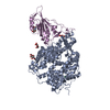
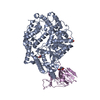
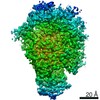
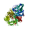
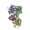
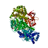
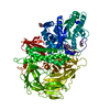
Journal: Nat Microbiol / Year: 2021 Title: SARS-CoV-2 variant prediction and antiviral drug design are enabled by RBD in vitro evolution. Authors: Jiří Zahradník / Shir Marciano / Maya Shemesh / Eyal Zoler / Daniel Harari / Jeanne Chiaravalli / Björn Meyer / Yinon Rudich / Chunlin Li / Ira Marton / Orly Dym / Nadav Elad / Mark G ...Authors: Jiří Zahradník / Shir Marciano / Maya Shemesh / Eyal Zoler / Daniel Harari / Jeanne Chiaravalli / Björn Meyer / Yinon Rudich / Chunlin Li / Ira Marton / Orly Dym / Nadav Elad / Mark G Lewis / Hanne Andersen / Matthew Gagne / Robert A Seder / Daniel C Douek / Gideon Schreiber / Abstract: SARS-CoV-2 variants of interest and concern will continue to emerge for the duration of the COVID-19 pandemic. To map mutations in the receptor-binding domain (RBD) of the spike protein that affect ...SARS-CoV-2 variants of interest and concern will continue to emerge for the duration of the COVID-19 pandemic. To map mutations in the receptor-binding domain (RBD) of the spike protein that affect binding to angiotensin-converting enzyme 2 (ACE2), the receptor for SARS-CoV-2, we applied in vitro evolution to affinity-mature the RBD. Multiple rounds of random mutagenic libraries of the RBD were sorted against decreasing concentrations of ACE2, resulting in the selection of higher affinity RBD binders. We found that mutations present in more transmissible viruses (S477N, E484K and N501Y) were preferentially selected in our high-throughput screen. Evolved RBD mutants include prominently the amino acid substitutions found in the RBDs of B.1.620, B.1.1.7 (Alpha), B1.351 (Beta) and P.1 (Gamma) variants. Moreover, the incidence of RBD mutations in the population as presented in the GISAID database (April 2021) is positively correlated with increased binding affinity to ACE2. Further in vitro evolution increased binding by 1,000-fold and identified mutations that may be more infectious if they evolve in the circulating viral population, for example, Q498R is epistatic to N501Y. We show that our high-affinity variant RBD-62 can be used as a drug to inhibit infection with SARS-CoV-2 and variants Alpha, Beta and Gamma in vitro. In a model of SARS-CoV-2 challenge in hamster, RBD-62 significantly reduced clinical disease when administered before or after infection. A 2.9 Å cryo-electron microscopy structure of the high-affinity complex of RBD-62 and ACE2, including all rapidly spreading mutations, provides a structural basis for future drug and vaccine development and for in silico evaluation of known antibodies.
History
Deposition
Jan 11, 2021
-
Header (metadata) release
Feb 17, 2021
-
Map release
Feb 17, 2021
-
Update
Sep 22, 2021
-
Current status
Sep 22, 2021
Processing site: PDBe / Status: Released
-
Structure visualization
Movie
Surface view with section colored by density value
Model: UltrAuFoil R1.2/1.3 / Material: GOLD / Mesh: 300 / Support film - Material: GOLD / Support film - topology: HOLEY / Pretreatment - Type: GLOW DISCHARGE / Pretreatment - Atmosphere: AIR
Vitrification
Cryogen name: ETHANE / Chamber humidity: 100 % / Chamber temperature: 277 K / Instrument: FEI VITROBOT MARK IV
-
Electron microscopy
Microscope
TFS KRIOS
Image recording
Film or detector model: GATAN K3 (6k x 4k) / Number grids imaged: 2 / Number real images: 4470 / Average exposure time: 1.214 sec. / Average electron dose: 70.0 e/Å2
Electron beam
Acceleration voltage: 300 kV / Electron source: FIELD EMISSION GUN
In the structure databanks used in Yorodumi, some data are registered as the other names, "COVID-19 virus" and "2019-nCoV". Here are the details of the virus and the list of structure data.
Jan 31, 2019. EMDB accession codes are about to change! (news from PDBe EMDB page)
EMDB accession codes are about to change! (news from PDBe EMDB page)
The allocation of 4 digits for EMDB accession codes will soon come to an end. Whilst these codes will remain in use, new EMDB accession codes will include an additional digit and will expand incrementally as the available range of codes is exhausted. The current 4-digit format prefixed with “EMD-” (i.e. EMD-XXXX) will advance to a 5-digit format (i.e. EMD-XXXXX), and so on. It is currently estimated that the 4-digit codes will be depleted around Spring 2019, at which point the 5-digit format will come into force.
The EM Navigator/Yorodumi systems omit the EMD- prefix.
Related info.:Q: What is EMD? / ID/Accession-code notation in Yorodumi/EM Navigator
Yorodumi is a browser for structure data from EMDB, PDB, SASBDB, etc.
This page is also the successor to EM Navigator detail page, and also detail information page/front-end page for Omokage search.
The word "yorodu" (or yorozu) is an old Japanese word meaning "ten thousand". "mi" (miru) is to see.
Related info.:EMDB / PDB / SASBDB / Comparison of 3 databanks / Yorodumi Search / Aug 31, 2016. New EM Navigator & Yorodumi / Yorodumi Papers / Jmol/JSmol / Function and homology information / Changes in new EM Navigator and Yorodumi
 Movie
Movie Controller


 Open data
 Basic information
 Map data
 Sample
 Function and homology information
 Homo sapiens (human) /
 Severe acute respiratory syndrome coronavirus 2
 Authors
 Israel, 2 items
  Citation

 Structure visualization
 Downloads & links
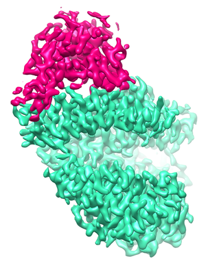 emd_12187.png
 http://ftp.pdbj.org/pub/emdb/structures/EMD-12187


 Z
Z Y
Y X
X


 Sample components
 Processing
 Electron microscopy
 FIELD EMISSION GUN