Movie
Movie Controller

[English] 日本語
 Yorodumi
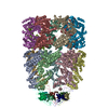
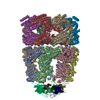
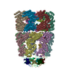
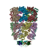
- EMDB-1180: Allosteric signaling of ATP hydrolysis in GroEL-GroES complexes. -
+ Open data

- Basic information
| Entry | Database: EMDB / ID: EMD-1180 | |||||||||
|---|---|---|---|---|---|---|---|---|---|---|
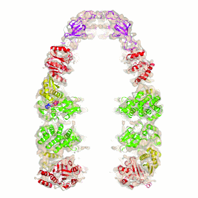
| Title | Allosteric signaling of ATP hydrolysis in GroEL-GroES complexes. | |||||||||
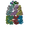
 Map data Map data | GroEL-ATP7-GroES complex | |||||||||
 Sample Sample |
| |||||||||
| Function / homology |  Function and homology information Function and homology informationGroEL-GroES complex / chaperonin ATPase / virion assembly / isomerase activity / protein folding chaperone / ATP-dependent protein folding chaperone / response to radiation / : / response to heat / protein refolding ...GroEL-GroES complex / chaperonin ATPase / virion assembly / isomerase activity / protein folding chaperone / ATP-dependent protein folding chaperone / response to radiation / : / response to heat / protein refolding / protein-folding chaperone binding / protein folding / magnesium ion binding / ATP hydrolysis activity / ATP binding / membrane / metal ion binding / identical protein binding / cytosol Similarity search - Function | |||||||||
| Biological species |  | |||||||||
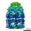
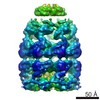
| Method | single particle reconstruction / cryo EM / Resolution: 7.7 Å | |||||||||
 Authors Authors | Ranson NA | |||||||||
 Citation Citation | Journal: Nat Struct Mol Biol / Year: 2006 Title: Allosteric signaling of ATP hydrolysis in GroEL-GroES complexes. Authors: Neil A Ranson / Daniel K Clare / George W Farr / David Houldershaw / Arthur L Horwich / Helen R Saibil /  Abstract: The double-ring chaperonin GroEL and its lid-like cochaperonin GroES form asymmetric complexes that, in the ATP-bound state, mediate productive folding in a hydrophilic, GroES-encapsulated chamber, ...The double-ring chaperonin GroEL and its lid-like cochaperonin GroES form asymmetric complexes that, in the ATP-bound state, mediate productive folding in a hydrophilic, GroES-encapsulated chamber, the so-called cis cavity. Upon ATP hydrolysis within the cis ring, the asymmetric complex becomes able to accept non-native polypeptides and ATP in the open, trans ring. Here we have examined the structural basis for this allosteric switch in activity by cryo-EM and single-particle image processing. ATP hydrolysis does not change the conformation of the cis ring, but its effects are transmitted through an inter-ring contact and cause domain rotations in the mobile trans ring. These rigid-body movements in the trans ring lead to disruption of its intra-ring contacts, expansion of the entire ring and opening of both the nucleotide pocket and the substrate-binding domains, admitting ATP and new substrate protein. | |||||||||
| History |
|
- Structure visualization
| Movie |
Movie viewer |
|---|---|
| Structure viewer | EM map: SurfViewMolmilJmol/JSmol |
| Supplemental images |
- Downloads & links
-EMDB archive
| Map data | emd_1180.map.gz | 1.4 MB | EMDB map data format | |
|---|---|---|---|---|
| Header (meta data) | emd-1180-v30.xmlemd-1180.xml | 10.2 KB 10.2 KB | Display Display | EMDB header |
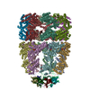
| Images |  1180.gif 1180.gif | 38.3 KB | ||
| Archive directory |  http://ftp.pdbj.org/pub/emdb/structures/EMD-1180ftp://ftp.pdbj.org/pub/emdb/structures/EMD-1180 http://ftp.pdbj.org/pub/emdb/structures/EMD-1180ftp://ftp.pdbj.org/pub/emdb/structures/EMD-1180 | HTTPS FTP |
-Related structure data
| Related structure data | 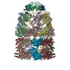 2c7cMC 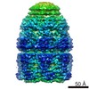 1181C 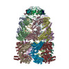 2c7dC C: citing same article ( M: atomic model generated by this map |
|---|---|
| Similar structure data |
-Links
| EMDB pages | EMDB (EBI/PDBe) / EMDataResource |
|---|---|
| Related items in Molecule of the Month |


-Map
| File | Download / File: emd_1180.map.gz / Format: CCP4 / Size: 26.4 MB / Type: IMAGE STORED AS FLOATING POINT NUMBER (4 BYTES) | ||||||||||||||||||||||||||||||||||||||||||||||||||||||||||||||||||||
|---|---|---|---|---|---|---|---|---|---|---|---|---|---|---|---|---|---|---|---|---|---|---|---|---|---|---|---|---|---|---|---|---|---|---|---|---|---|---|---|---|---|---|---|---|---|---|---|---|---|---|---|---|---|---|---|---|---|---|---|---|---|---|---|---|---|---|---|---|---|
| Annotation | GroEL-ATP7-GroES complex | ||||||||||||||||||||||||||||||||||||||||||||||||||||||||||||||||||||
| Projections & slices | Image control
Images are generated by Spider. | ||||||||||||||||||||||||||||||||||||||||||||||||||||||||||||||||||||
| Voxel size | X=Y=Z: 1.4 Å | ||||||||||||||||||||||||||||||||||||||||||||||||||||||||||||||||||||
| Density |
| ||||||||||||||||||||||||||||||||||||||||||||||||||||||||||||||||||||
| Symmetry | Space group: 1 | ||||||||||||||||||||||||||||||||||||||||||||||||||||||||||||||||||||
| Details | EMDB XML:
CCP4 map header:
| ||||||||||||||||||||||||||||||||||||||||||||||||||||||||||||||||||||
 Z (Sec.)
 X (Row.)
 Y (Col.)


-Supplemental data
- Sample components
-Entire : GroEL-ATP7-GroES
| Entire | Name: GroEL-ATP7-GroES |
|---|---|
| Components |
|
-Supramolecule #1000: GroEL-ATP7-GroES
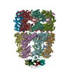
| Supramolecule | Name: GroEL-ATP7-GroES / type: sample / ID: 1000 Oligomeric state: One tetradecamer of GroEL plus one heptamer of GroES Number unique components: 2 |
|---|---|
| Molecular weight | Experimental: 900 KDa / Theoretical: 900 KDa |
-Macromolecule #1: GroEL
| Macromolecule | Name: GroEL / type: protein_or_peptide / ID: 1 / Details: GroEL D398A / Number of copies: 14 / Oligomeric state: tetradecamer / Recombinant expression: Yes |
|---|---|
| Source (natural) | Organism: |
| Recombinant expression | Organism: |
-Macromolecule #2: GroES
| Macromolecule | Name: GroES / type: protein_or_peptide / ID: 2 / Number of copies: 7 / Oligomeric state: heptamer / Recombinant expression: Yes |
|---|---|
| Source (natural) | Organism: |
| Recombinant expression | Organism: |
-Experimental details
-Structure determination
| Method | cryo EM |
|---|---|
 Processing Processing | single particle reconstruction |
| Aggregation state | particle |
-Sample preparation
| Concentration | 1.0 mg/mL |
|---|---|
| Buffer | pH: 7.5 / Details: 12.5 mM HEPES, ph7.5 5mM KCl 5mM MgCl2 |
| Grid | Details: 300 mesh Cu grid - holey carbon |
| Vitrification | Cryogen name: ETHANE / Chamber humidity: 95 % / Chamber temperature: 100 K / Timed resolved state: Vitrified within 30s of mixing / Method: blot for 3-4s before plunging |
- Electron microscopy
| Microscope | FEI TECNAI F20 |
|---|---|
| Image recording | Category: FILM / Film or detector model: KODAK SO-163 FILM / Digitization - Scanner: ZEISS SCAI / Digitization - Sampling interval: 7 µm / Number real images: 189 / Average electron dose: 15 e/Å2 / Bits/pixel: 8 |
| Tilt angle min | 0 |
| Tilt angle max | 0 |
| Electron beam | Acceleration voltage: 200 kV / Electron source:  FIELD EMISSION GUN FIELD EMISSION GUN |
| Electron optics | Calibrated magnification: 50000 / Illumination mode: FLOOD BEAM / Imaging mode: BRIGHT FIELD / Cs: 2.0 mm / Nominal defocus max: 3.2 µm / Nominal defocus min: 1.1 µm / Nominal magnification: 50000 |
| Sample stage | Specimen holder: single tilt cryo / Specimen holder model: GATAN LIQUID NITROGEN |
| Experimental equipment |  Model: Tecnai F20 / Image courtesy: FEI Company |
-Image processing
| Details | manual particle selection |
|---|---|
| CTF correction | Details: full correction on 2D class averages |
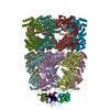
| Final reconstruction | Applied symmetry - Point group: C7 (7 fold cyclic) / Algorithm: OTHER / Resolution.type: BY AUTHOR / Resolution: 7.7 Å / Resolution method: FSC 0.5 CUT-OFF / Software - Name: SPIDER / Number images used: 16281 |
-Atomic model buiding 1
| Initial model | PDB ID: |
|---|---|
| Software | Name: URO |
| Details | Protocol: rigid body. Seven independent domains (equatorial, intermediate and apical domains of each GroEL ring, plus GroES) |
| Refinement | Space: RECIPROCAL / Protocol: RIGID BODY FIT |
| Output model | PDB-2c7c: |