Movie
Movie Controller

[English] 日本語
 Yorodumi
- EMDB-1531: Three-dimensional structure of Aquifex aeolicus co-chaperonin pro... -
+ Open data

- Basic information
| Entry | Database: EMDB / ID: EMD-1531 | |||||||||
|---|---|---|---|---|---|---|---|---|---|---|
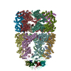
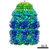
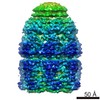
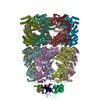
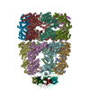
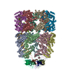
| Title | Three-dimensional structure of Aquifex aeolicus co-chaperonin protein 10 complexed with GroEL and ADP by cryo-EM | |||||||||
 Map data Map data | none | |||||||||
 Sample Sample |
| |||||||||
 Keywords Keywords | co-chaperonin / hyper-thermophile / conformational heterogeneity / electron cryomicroscopy | |||||||||
| Function / homology | :  Function and homology information Function and homology information | |||||||||
| Biological species |   Aquifex aeolicus (bacteria) Aquifex aeolicus (bacteria) | |||||||||
| Method | single particle reconstruction / cryo EM / negative staining / Resolution: 8.0 Å | |||||||||
 Authors Authors | Chen DH / Luke K / Zhang J / Chiu W / Wittung-Stafshede P | |||||||||
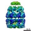
 Citation Citation | Journal: J Mol Biol / Year: 2008 Title: Location and flexibility of the unique C-terminal tail of Aquifex aeolicus co-chaperonin protein 10 as derived by cryo-electron microscopy and biophysical techniques. Authors: Dong-Hua Chen / Kathryn Luke / Junjie Zhang / Wah Chiu / Pernilla Wittung-Stafshede /  Abstract: Co-chaperonin protein 10 (cpn10, GroES in Escherichia coli) is a ring-shaped heptameric protein that facilitates substrate folding when in complex with cpn60 (GroEL in E. coli). The cpn10 from the ...Co-chaperonin protein 10 (cpn10, GroES in Escherichia coli) is a ring-shaped heptameric protein that facilitates substrate folding when in complex with cpn60 (GroEL in E. coli). The cpn10 from the hyperthermophilic, ancient bacterium Aquifex aeolicus (Aacpn10) has a 25-residue C-terminal extension in each monomer not found in any other cpn10 protein. Earlier in vitro work has shown that this tail is not needed for heptamer assembly or protein function. Without the tail, however, the heptamers (Aacpn10del-25) readily aggregate into fibrillar stacked rings. To explain this phenomenon, we performed binding experiments with a peptide construct of the tail to establish its specificity for Aacpn10del-25 and used cryo-electron microscopy to determine the three-dimensional (3D) structure of the GroEL-Aacpn10-ADP complex at an 8-A resolution. We found that the GroEL-Aacpn10 structure is similar to the GroEL-GroES structure at this resolution, suggesting that Aacpn10 has molecular interactions with cpn60 similar to other cpn10s. The cryo-electron microscopy density map does not directly reveal the density of the Aacpn10 25-residue tail. However, the 3D statistical variance coefficient map computed from multiple 3D reconstructions with randomly selected particle images suggests that the tail is located at the Aacpn10 monomer-monomer interface and extends toward the cis-ring apical domain of GroEL. The tail at this location does not block the formation of a functional co-chaperonin/chaperonin complex but limits self-aggregation into linear fibrils at high temperatures. In addition, the 3D variance coefficient map identifies several regions inside the GroEL-Aacpn10 complex that have flexible conformations. This observation is in full agreement with the structural properties of an effective chaperonin. | |||||||||
| History |
|
- Structure visualization
| Movie |
Movie viewer |
|---|---|
| Structure viewer | EM map: SurfViewMolmilJmol/JSmol |
| Supplemental images |

- Downloads & links
-EMDB archive
| Map data | emd_1531.map.gz | 2.6 MB | EMDB map data format | |
|---|---|---|---|---|
| Header (meta data) | emd-1531-v30.xmlemd-1531.xml | 12.5 KB 12.5 KB | Display Display | EMDB header |
| Images |  1531.gif 1531.gif | 218.5 KB | ||
| Archive directory |  http://ftp.pdbj.org/pub/emdb/structures/EMD-1531ftp://ftp.pdbj.org/pub/emdb/structures/EMD-1531 http://ftp.pdbj.org/pub/emdb/structures/EMD-1531ftp://ftp.pdbj.org/pub/emdb/structures/EMD-1531 | HTTPS FTP |
-Related structure data
| Similar structure data |
|---|

-Links
| EMDB pages | EMDB (EBI/PDBe) / EMDataResource |
|---|
-Map
| File | Download / File: emd_1531.map.gz / Format: CCP4 / Size: 29.8 MB / Type: IMAGE STORED AS FLOATING POINT NUMBER (4 BYTES) | ||||||||||||||||||||||||||||||||||||||||||||||||||||||||||||||||||||
|---|---|---|---|---|---|---|---|---|---|---|---|---|---|---|---|---|---|---|---|---|---|---|---|---|---|---|---|---|---|---|---|---|---|---|---|---|---|---|---|---|---|---|---|---|---|---|---|---|---|---|---|---|---|---|---|---|---|---|---|---|---|---|---|---|---|---|---|---|---|
| Annotation | none | ||||||||||||||||||||||||||||||||||||||||||||||||||||||||||||||||||||
| Projections & slices | Image control
Images are generated by Spider. | ||||||||||||||||||||||||||||||||||||||||||||||||||||||||||||||||||||
| Voxel size | X=Y=Z: 1.795 Å | ||||||||||||||||||||||||||||||||||||||||||||||||||||||||||||||||||||
| Density |
| ||||||||||||||||||||||||||||||||||||||||||||||||||||||||||||||||||||
| Symmetry | Space group: 1 | ||||||||||||||||||||||||||||||||||||||||||||||||||||||||||||||||||||
| Details | EMDB XML:
CCP4 map header:
| ||||||||||||||||||||||||||||||||||||||||||||||||||||||||||||||||||||
 Z (Sec.)
 Y (Row.)
 X (Col.)


-Supplemental data
- Sample components
-Entire : Aacpn10 capped to one end of GroEL under Mg-ADP
| Entire | Name: Aacpn10 capped to one end of GroEL under Mg-ADP |
|---|---|
| Components |
|
-Supramolecule #1000: Aacpn10 capped to one end of GroEL under Mg-ADP
| Supramolecule | Name: Aacpn10 capped to one end of GroEL under Mg-ADP / type: sample / ID: 1000 Details: Aacpn10 was incubated with GroEL in 50 mM Tris-HCl, 30 mM MgCl2, 2 mM ADP, pH 7.5 at 37 Celsius for 1 hour Oligomeric state: One heptamer of Aacpn10 and seven ADP bind to one heptamer of GroEL Number unique components: 2 |
|---|---|
| Molecular weight | Experimental: 900 KDa / Theoretical: 900 KDa |
-Macromolecule #1: Chaperonin
| Macromolecule | Name: Chaperonin / type: protein_or_peptide / ID: 1 / Name.synonym: GroEL / Number of copies: 1 / Oligomeric state: heptamer / Recombinant expression: Yes |
|---|---|
| Source (natural) | Organism: |
| Molecular weight | Experimental: 800 KDa / Theoretical: 800 KDa |
| Recombinant expression | Organism: |
| Sequence | InterPro: INTERPRO: IPR012723 |
-Macromolecule #2: Aquifex aeolicus co-chaperonin 10
| Macromolecule | Name: Aquifex aeolicus co-chaperonin 10 / type: protein_or_peptide / ID: 2 / Name.synonym: Aacpn10 / Number of copies: 1 / Oligomeric state: heptamer / Recombinant expression: Yes |
|---|---|
| Source (natural) | Organism: Aquifex aeolicus (bacteria) / Strain: AQ2199 / Tissue: Bacterial / Cell: Aquifex aeolicus / Location in cell: Cytosol |
| Molecular weight | Experimental: 100 KDa / Theoretical: 100 KDa |
| Recombinant expression | Organism: |
-Experimental details
-Structure determination
| Method | negative staining, cryo EM |
|---|---|
 Processing Processing | single particle reconstruction |
| Aggregation state | particle |
-Sample preparation
| Concentration | 4 mg/mL |
|---|---|
| Buffer | pH: 7.5 / Details: 50 mM Tris-HCl, 30 mM MgCl2 |
| Staining | Type: NEGATIVE Details: The sample solution was blotted for 2.5 s before submersion in liquid ethane cooled by liquid nitrogen using FEI Vitrobot |
| Grid | Details: 400 mesh copper grid (R1.2/1.3 Quantifoil) |
| Vitrification | Cryogen name: ETHANE / Chamber humidity: 95 % / Chamber temperature: 4.2 K / Instrument: OTHER / Details: Vitrification instrument: Vitrobot / Method: blot for 2.5 s before plunging |
- Electron microscopy
| Microscope | JEOL 3000SFF |
|---|---|
| Temperature | Min: 4.2 K / Max: 4.2 K / Average: 4.2 K |
| Alignment procedure | Legacy - Astigmatism: objective lens astigmatism was corrected at 400,000 times magnification |
| Details | Yoshi MDS box for low-dose imaging |
| Date | Feb 16, 2007 |
| Image recording | Category: FILM / Film or detector model: KODAK SO-163 FILM / Digitization - Scanner: NIKON SUPER COOLSCAN 9000 / Digitization - Sampling interval: 6.35 µm / Number real images: 720 / Average electron dose: 36 e/Å2 Details: The particle images were averaged by 2 to give 1.91 Angstrom per pixel. The final corrected pixel size for the 3D density map is 1.8 Angstrom per pixel. Bits/pixel: 8 |
| Electron beam | Acceleration voltage: 300 kV / Electron source:  FIELD EMISSION GUN FIELD EMISSION GUN |
| Electron optics | Illumination mode: FLOOD BEAM / Imaging mode: BRIGHT FIELD / Cs: 1.6 mm / Nominal defocus max: 3.4 µm / Nominal defocus min: 0.86 µm / Nominal magnification: 50000 |
| Sample stage | Specimen holder: top-entry / Specimen holder model: OTHER |
+Image processing
-Atomic model buiding 1
| Initial model | PDB ID: Chain - Chain ID: A |
|---|---|
| Software | Name: UROX |
| Details | PDBEntryID_givenInChain. Protocol: domain-based rigid body. UROX was used to perform the domain-as-rigid-body fitting of the GroEL-GroES-ADP crystal structure into the cryo-EM density map of the GroEL-Aacpn10-ADP complex |
| Refinement | Space: REAL / Protocol: RIGID BODY FIT / Target criteria: cross correlation |