[English] 日本語
Yorodumi
- EMDB-10263: Structure of Coxsackievirus A10 complexed with its receptor KREMEN1 -

+
Open data


ID or keywords:

Loading...

-
Basic information

Entry
Database: EMDB / ID: EMD-10263
TitleStructure of Coxsackievirus A10 complexed with its receptor KREMEN1
Map data
Sample
  • Complex: Coxsackievirus A10
    • Complex: Coxsackievirus A10
      • Protein or peptide: Capsid protein VP1
      • Protein or peptide: Coxsackievirus VP2
      • Protein or peptide: Capsid protein VP3
      • Protein or peptide: Coxsackievirus VP4
    • Complex: KREMEN1
      • Protein or peptide: Kremen protein 1
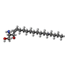
  • Ligand: SPHINGOSINE
  • Ligand: 2-acetamido-2-deoxy-beta-D-glucopyranose
KeywordsCV-A10 / KREMEN1 / Virus-receptor complex / Hand / foot and mouth disease / VIRUS / picornavirus uncoating intermediate
Function / homology
Function and homology information


Signaling by LRP5 mutants / Negative regulation of TCF-dependent signaling by WNT ligand antagonists / negative regulation of axon regeneration / cell communication / negative regulation of ossification / regulation of canonical Wnt signaling pathway / limb development / symbiont-mediated suppression of host cytoplasmic pattern recognition receptor signaling pathway via inhibition of MDA-5 activity / picornain 2A / symbiont-mediated suppression of host mRNA export from nucleus ...Signaling by LRP5 mutants / Negative regulation of TCF-dependent signaling by WNT ligand antagonists / negative regulation of axon regeneration / cell communication / negative regulation of ossification / regulation of canonical Wnt signaling pathway / limb development / symbiont-mediated suppression of host cytoplasmic pattern recognition receptor signaling pathway via inhibition of MDA-5 activity / picornain 2A / symbiont-mediated suppression of host mRNA export from nucleus / symbiont genome entry into host cell via pore formation in plasma membrane / picornain 3C / T=pseudo3 icosahedral viral capsid / TCF dependent signaling in response to WNT / host cell cytoplasmic vesicle membrane / negative regulation of canonical Wnt signaling pathway / cytoplasmic vesicle membrane / endocytosis involved in viral entry into host cell / Wnt signaling pathway / nucleoside-triphosphate phosphatase / channel activity / monoatomic ion transmembrane transport / RNA helicase activity / induction by virus of host autophagy / RNA-directed RNA polymerase / viral RNA genome replication / cysteine-type endopeptidase activity / RNA-dependent RNA polymerase activity / virus-mediated perturbation of host defense response / neuronal cell body / DNA-templated transcription / host cell nucleus / virion attachment to host cell / apoptotic process / structural molecule activity / ATP hydrolysis activity / proteolysis / RNA binding / ATP binding / membrane / metal ion binding / plasma membrane
Similarity search - Function
Kremen / : / Carbohydrate-binding WSC / WSC domain / WSC domain profile. / present in yeast cell wall integrity and stress response component proteins / CUB domain / Domain first found in C1r, C1s, uEGF, and bone morphogenetic protein. / CUB domain / CUB domain profile. ...Kremen / : / Carbohydrate-binding WSC / WSC domain / WSC domain profile. / present in yeast cell wall integrity and stress response component proteins / CUB domain / Domain first found in C1r, C1s, uEGF, and bone morphogenetic protein. / CUB domain / CUB domain profile. / Spermadhesin, CUB domain superfamily / Picornavirus coat protein / Kringle domain / Kringle / Kringle, conserved site / Kringle superfamily / Kringle domain signature. / Kringle domain profile. / Kringle domain / Kringle-like fold / Poliovirus 3A protein-like / Poliovirus 3A protein like / Picornavirus 2B protein / Poliovirus core protein 3a, soluble domain / Picornavirus 2B protein / Peptidase C3, picornavirus core protein 2A / Picornavirus core protein 2A / Picornavirus coat protein VP4 / Picornavirus coat protein (VP4) / Peptidase C3A/C3B, picornaviral / 3C cysteine protease (picornain 3C) / Picornavirales 3C/3C-like protease domain / Picornavirales 3C/3C-like protease domain profile. / Picornavirus capsid / picornavirus capsid protein / Helicase, superfamily 3, single-stranded RNA virus / Superfamily 3 helicase of positive ssRNA viruses domain profile. / Helicase, superfamily 3, single-stranded DNA/RNA virus / RNA helicase / Picornavirus/Calicivirus coat protein / Viral coat protein subunit / RNA-directed RNA polymerase, C-terminal domain / Viral RNA-dependent RNA polymerase / Reverse transcriptase/Diguanylate cyclase domain / RNA-directed RNA polymerase, catalytic domain / RdRp of positive ssRNA viruses catalytic domain profile. / ATPases associated with a variety of cellular activities / AAA+ ATPase domain / Peptidase S1, PA clan, chymotrypsin-like fold / Peptidase S1, PA clan / DNA/RNA polymerase superfamily / P-loop containing nucleoside triphosphate hydrolase
Similarity search - Domain/homology
Genome polyprotein / Kremen protein 1
Similarity search - Component
Biological speciesCoxsackievirus A10 / Homo sapiens (human)
Methodsingle particle reconstruction / cryo EM / Resolution: 3.9 Å
AuthorsZhao Y / Zhou D
Funding support United Kingdom, 3 items
OrganizationGrant numberCountry
Medical Research Council (United Kingdom)MR/N00065X/1 United Kingdom
Wellcome Trust101122/Z/13/Z United Kingdom
Cancer Research UKC375/A17721 United Kingdom
CitationJournal: Nat Commun / Year: 2020
Title: Hand-foot-and-mouth disease virus receptor KREMEN1 binds the canyon of Coxsackie Virus A10.
Authors: Yuguang Zhao / Daming Zhou / Tao Ni / Dimple Karia / Abhay Kotecha / Xiangxi Wang / Zihe Rao / E Yvonne Jones / Elizabeth E Fry / Jingshan Ren / David I Stuart /
Abstract: Coxsackievirus A10 (CV-A10) is responsible for an escalating number of severe infections in children, but no prophylactics or therapeutics are currently available. KREMEN1 (KRM1) is the entry ...Coxsackievirus A10 (CV-A10) is responsible for an escalating number of severe infections in children, but no prophylactics or therapeutics are currently available. KREMEN1 (KRM1) is the entry receptor for the largest receptor-group of hand-foot-and-mouth disease causing viruses, which includes CV-A10. We report here structures of CV-A10 mature virus alone and in complex with KRM1 as well as of the CV-A10 A-particle. The receptor spans the viral canyon with a large footprint on the virus surface. The footprint has some overlap with that seen for the neonatal Fc receptor complexed with enterovirus E6 but is larger and distinct from that of another enterovirus receptor SCARB2. Reduced occupancy of a particle-stabilising pocket factor in the complexed virus and the presence of both unbound and expanded virus particles suggests receptor binding initiates a cascade of conformational changes that produces expanded particles primed for viral uncoating.
History
DepositionAug 27, 2019-
Header (metadata) releaseSep 4, 2019-
Map releaseJan 15, 2020-
UpdateNov 6, 2024-
Current statusNov 6, 2024Processing site: PDBe / Status: Released

-
Structure visualization

Movie
  • Surface view with section colored by density value
  • Surface level: 0.01
  • Imaged by UCSF Chimera
  • Download
  • Surface view colored by radius
  • Surface level: 0.01
  • Imaged by UCSF Chimera
  • Download
  • Surface view with fitted model
  • Atomic models: PDB-6snw
  • Surface level: 0.01
  • Imaged by UCSF Chimera
  • Download
  • Simplified surface model + fitted atomic model
  • Atomic modelsPDB-6snw
  • Imaged by Jmol
  • Download
Movie viewer
Structure viewerEM map:
SurfViewMolmilJmol/JSmol
Supplemental images

Downloads & links

-
Map

FileDownload / File: emd_10263.map.gz / Format: CCP4 / Size: 244.1 MB / Type: IMAGE STORED AS FLOATING POINT NUMBER (4 BYTES)
Projections & slices

Image control

Size
Brightness
Contrast
Others
AxesZ (Sec.)Y (Row.)X (Col.)
1.35 Å/pix.
x 400 pix.
= 540. Å
1.35 Å/pix.
x 400 pix.
= 540. Å
1.35 Å/pix.
x 400 pix.
= 540. Å

Surface

Projections

Slices (1/3)

Slices (1/2)

Slices (2/3)

Images are generated by Spider.

Voxel sizeX=Y=Z: 1.35 Å
Density
Contour LevelBy AUTHOR: 0.01 / Movie #1: 0.01
Minimum - Maximum-0.05129708 - 0.097188205
Average (Standard dev.)0.0006619525 (±0.0049746973)
SymmetrySpace group: 1
Details

EMDB XML:

Map geometry
Axis orderXYZ
Origin000
Dimensions400400400
Spacing400400400
CellA=B=C: 540.0 Å
α=β=γ: 90.0 °

CCP4 map header:

modeImage stored as Reals
Å/pix. X/Y/Z1.351.351.35
M x/y/z400400400
origin x/y/z0.0000.0000.000
length x/y/z540.000540.000540.000
α/β/γ90.00090.00090.000
MAP C/R/S123
start NC/NR/NS000
NC/NR/NS400400400
D min/max/mean-0.0510.0970.001

-
Supplemental data

-
Sample components

+
Entire : Coxsackievirus A10

EntireName: Coxsackievirus A10
Components
  • Complex: Coxsackievirus A10
    • Complex: Coxsackievirus A10
      • Protein or peptide: Capsid protein VP1
      • Protein or peptide: Coxsackievirus VP2
      • Protein or peptide: Capsid protein VP3
      • Protein or peptide: Coxsackievirus VP4
    • Complex: KREMEN1
      • Protein or peptide: Kremen protein 1
  • Ligand: SPHINGOSINE
  • Ligand: 2-acetamido-2-deoxy-beta-D-glucopyranose

+
Supramolecule #1: Coxsackievirus A10

SupramoleculeName: Coxsackievirus A10 / type: complex / ID: 1 / Parent: 0 / Macromolecule list: #1-#5

+
Supramolecule #2: Coxsackievirus A10

SupramoleculeName: Coxsackievirus A10 / type: complex / ID: 2 / Parent: 1 / Macromolecule list: #1-#4
Source (natural)Organism: Coxsackievirus A10 / Strain: Kowalik

+
Supramolecule #3: KREMEN1

SupramoleculeName: KREMEN1 / type: complex / ID: 3 / Parent: 1 / Macromolecule list: #5
Source (natural)Organism: Homo sapiens (human)

+
Macromolecule #1: Capsid protein VP1

MacromoleculeName: Capsid protein VP1 / type: protein_or_peptide / ID: 1 / Number of copies: 1 / Enantiomer: LEVO / EC number: picornain 2A
Source (natural)Organism: Coxsackievirus A10
Molecular weightTheoretical: 33.191422 KDa
SequenceString: GDPVEDIIHD ALSSTVRRAI TSGQDVNTAA GTAPSSHRLE TGRVPALQAA ETGATSNATD ENMIETRCVM NRNGVLEATI SHFFSRSGL VGVVNLTDGG TDTTGYAVWD IDIMGFVQLR RKCEMFTYMR FNAEFTFVTT TENGEARPFM LQYMYVPPGA P KPTGRDAF ...String:
GDPVEDIIHD ALSSTVRRAI TSGQDVNTAA GTAPSSHRLE TGRVPALQAA ETGATSNATD ENMIETRCVM NRNGVLEATI SHFFSRSGL VGVVNLTDGG TDTTGYAVWD IDIMGFVQLR RKCEMFTYMR FNAEFTFVTT TENGEARPFM LQYMYVPPGA P KPTGRDAF QWQTATNPSV FVKLTDPPAQ VSVPFMSPAS AYQWFYDGYP TFGQHPETSN TTYGQCPNNM MGTFAVRVVS RV ASQLKLQ TRVYMKLKHV RAWIPRPIRS QPYLLKNFPN YDSSKITYSA RDRASIKQAN M

UniProtKB: Genome polyprotein

+
Macromolecule #2: Coxsackievirus VP2

MacromoleculeName: Coxsackievirus VP2 / type: protein_or_peptide / ID: 2 / Number of copies: 1 / Enantiomer: LEVO / EC number: picornain 2A
Source (natural)Organism: Coxsackievirus A10
Molecular weightTheoretical: 27.72998 KDa
SequenceString: SPSVEACGYS DRVAQLTVGN SSITTQEAAN IVLAYGEWPE YCPDTDATAV DKPTRPDVSV NRFYTLDSKM WQENSTGWYW KFPDVLNKT GVFGQNAQFH YLYRSGFCLH VQCNASKFHQ GALLVAVIPE FVLAGRGSNT KPNEAPHPGF NTTFPGTAGA S FNDPYVLD ...String:
SPSVEACGYS DRVAQLTVGN SSITTQEAAN IVLAYGEWPE YCPDTDATAV DKPTRPDVSV NRFYTLDSKM WQENSTGWYW KFPDVLNKT GVFGQNAQFH YLYRSGFCLH VQCNASKFHQ GALLVAVIPE FVLAGRGSNT KPNEAPHPGF NTTFPGTAGA S FNDPYVLD SGVPLSQSLI YPHQWINLRT NNCATIIVPY INAVPFDSAI NHSNFGLIVV PVSPLKYSSG ATTAIPITVT IA PLNSEFG GLRQAVSQ

UniProtKB: Genome polyprotein

+
Macromolecule #3: Capsid protein VP3

MacromoleculeName: Capsid protein VP3 / type: protein_or_peptide / ID: 3 / Number of copies: 1 / Enantiomer: LEVO / EC number: picornain 2A
Source (natural)Organism: Coxsackievirus A10
Molecular weightTheoretical: 26.219672 KDa
SequenceString: GLPTELRPGT NQFLTTEDDT AAPILPGFSP TPSIHIPGEV RSLLELCRVE TILEVNNTTD ATGLNRLLIP VSAQNKADEL CAAFMVDPG RIGPWQSTLV GQICRYYTQW SGSLKVTFMF TGSFMATGKM LIAYSPPGSA QPANRETAML GTHVIWDFGL Q SSVSLVIP ...String:
GLPTELRPGT NQFLTTEDDT AAPILPGFSP TPSIHIPGEV RSLLELCRVE TILEVNNTTD ATGLNRLLIP VSAQNKADEL CAAFMVDPG RIGPWQSTLV GQICRYYTQW SGSLKVTFMF TGSFMATGKM LIAYSPPGSA QPANRETAML GTHVIWDFGL Q SSVSLVIP WISNTHFRTA KTGGNYDYYT AGVVTLWYQT NYVVPPETPG EAYIIAMGAA QDNFTLKICK DTDEVTQQAV LQ

UniProtKB: Genome polyprotein

+
Macromolecule #4: Coxsackievirus VP4

MacromoleculeName: Coxsackievirus VP4 / type: protein_or_peptide / ID: 4 / Number of copies: 1 / Enantiomer: LEVO / EC number: picornain 2A
Source (natural)Organism: Coxsackievirus A10
Molecular weightTheoretical: 7.464065 KDa
SequenceString:
MGAQVSSQRS GSHETGNVAT GGSTINFTNI NYYKDSYAAS ASRQDFTQDP KKFTQPVLDS IRELSAPLN

UniProtKB: Genome polyprotein

+
Macromolecule #5: Kremen protein 1

MacromoleculeName: Kremen protein 1 / type: protein_or_peptide / ID: 5 / Number of copies: 1 / Enantiomer: LEVO
Source (natural)Organism: Homo sapiens (human)
Molecular weightTheoretical: 41.786293 KDa
Recombinant expressionOrganism: Homo sapiens (human)
SequenceString: ETGAPSPGLG PGPECFTANG ADYRGTQNWT ALQGGKPCLF WNETFQHPYN TLKYPNGEGG LGEHNYCRNP DGDVSPWCYV AEHEDGVYW KYCEIPACQM PGNLGCYKDH GNPPPLTGTS KTSNKLTIQT CISFCRSQRF KFAGMESGYA CFCGNNPDYW K YGEAASTE ...String:
ETGAPSPGLG PGPECFTANG ADYRGTQNWT ALQGGKPCLF WNETFQHPYN TLKYPNGEGG LGEHNYCRNP DGDVSPWCYV AEHEDGVYW KYCEIPACQM PGNLGCYKDH GNPPPLTGTS KTSNKLTIQT CISFCRSQRF KFAGMESGYA CFCGNNPDYW K YGEAASTE CNSVCFGDHT QPCGGDGRII LFDTLVGACG GNYSAMSSVV YSPDFPDTYA TGRVCYWTIR VPGASHIHFS FP LFDIRDS ADMVELLDGY THRVLARFHG RSRPPLSFNV SLDFVILYFF SDRINQAQGF AVLYQAVKEE GSENLYFQGG SLP QERPAV NQTVAEVITE QANLSVSAAR SSKVLYVITT SPSHPPQTVP GTHHHHHHHH HH

UniProtKB: Kremen protein 1

+
Macromolecule #6: SPHINGOSINE

MacromoleculeName: SPHINGOSINE / type: ligand / ID: 6 / Number of copies: 1 / Formula: SPH
Molecular weightTheoretical: 299.492 Da
Chemical component information

ChemComp-SPH:
SPHINGOSINE

+
Macromolecule #7: 2-acetamido-2-deoxy-beta-D-glucopyranose

MacromoleculeName: 2-acetamido-2-deoxy-beta-D-glucopyranose / type: ligand / ID: 7 / Number of copies: 2 / Formula: NAG
Molecular weightTheoretical: 221.208 Da
Chemical component information

ChemComp-NAG:
2-acetamido-2-deoxy-beta-D-glucopyranose

-
Experimental details

-
Structure determination

Methodcryo EM
Processingsingle particle reconstruction
Aggregation stateparticle

-
Sample preparation

BufferpH: 8
VitrificationCryogen name: ETHANE

-
Electron microscopy

MicroscopeFEI POLARA 300
Image recordingFilm or detector model: GATAN K2 SUMMIT (4k x 4k) / Number grids imaged: 1 / Average electron dose: 35.0 e/Å2
Electron beamAcceleration voltage: 300 kV / Electron source: FIELD EMISSION GUN
Electron opticsIllumination mode: FLOOD BEAM / Imaging mode: BRIGHT FIELD
Experimental equipment
Model: Tecnai Polara / Image courtesy: FEI Company

-
Image processing

Startup modelType of model: PDB ENTRY
PDB model - PDB ID:
Final reconstructionResolution.type: BY AUTHOR / Resolution: 3.9 Å / Resolution method: FSC 0.143 CUT-OFF / Number images used: 1597
Initial angle assignmentType: MAXIMUM LIKELIHOOD
Final angle assignmentType: MAXIMUM LIKELIHOOD

-
Atomic model buiding 1

RefinementSpace: REAL / Protocol: RIGID BODY FIT
Output model

PDB-6snw:
Structure of Coxsackievirus A10 complexed with its receptor KREMEN1

+
About Yorodumi

-
News

-
Feb 9, 2022. New format data for meta-information of EMDB entries

New format data for meta-information of EMDB entries

  • Version 3 of the EMDB header file is now the official format.
  • The previous official version 1.9 will be removed from the archive.

Related info.:EMDB header

External links:wwPDB to switch to version 3 of the EMDB data model

-
Aug 12, 2020. Covid-19 info

Covid-19 info

URL: https://pdbj.org/emnavi/covid19.php

New page: Covid-19 featured information page in EM Navigator.

Related info.:Covid-19 info / Mar 5, 2020. Novel coronavirus structure data

+
Mar 5, 2020. Novel coronavirus structure data

Novel coronavirus structure data

Related info.:Yorodumi Speices / Aug 12, 2020. Covid-19 info

External links:COVID-19 featured content - PDBj / Molecule of the Month (242):Coronavirus Proteases

+
Jan 31, 2019. EMDB accession codes are about to change! (news from PDBe EMDB page)

EMDB accession codes are about to change! (news from PDBe EMDB page)

  • The allocation of 4 digits for EMDB accession codes will soon come to an end. Whilst these codes will remain in use, new EMDB accession codes will include an additional digit and will expand incrementally as the available range of codes is exhausted. The current 4-digit format prefixed with “EMD-” (i.e. EMD-XXXX) will advance to a 5-digit format (i.e. EMD-XXXXX), and so on. It is currently estimated that the 4-digit codes will be depleted around Spring 2019, at which point the 5-digit format will come into force.
  • The EM Navigator/Yorodumi systems omit the EMD- prefix.

Related info.:Q: What is EMD? / ID/Accession-code notation in Yorodumi/EM Navigator

External links:EMDB Accession Codes are Changing Soon! / Contact to PDBj

+
Jul 12, 2017. Major update of PDB

Major update of PDB

  • wwPDB released updated PDB data conforming to the new PDBx/mmCIF dictionary.
  • This is a major update changing the version number from 4 to 5, and with Remediation, in which all the entries are updated.
  • In this update, many items about electron microscopy experimental information are reorganized (e.g. em_software).
  • Now, EM Navigator and Yorodumi are based on the updated data.

External links:wwPDB Remediation / Enriched Model Files Conforming to OneDep Data Standards Now Available in the PDB FTP Archive

-
Yorodumi

Thousand views of thousand structures

  • Yorodumi is a browser for structure data from EMDB, PDB, SASBDB, etc.
  • This page is also the successor to EM Navigator detail page, and also detail information page/front-end page for Omokage search.
  • The word "yorodu" (or yorozu) is an old Japanese word meaning "ten thousand". "mi" (miru) is to see.

Related info.:EMDB / PDB / SASBDB / Comparison of 3 databanks / Yorodumi Search / Aug 31, 2016. New EM Navigator & Yorodumi / Yorodumi Papers / Jmol/JSmol / Function and homology information / Changes in new EM Navigator and Yorodumi

Read more