 Movie
Movie Controller


+ Open data

- Basic information
| Entry | Database: EMDB / ID: EMD-0212 | |||||||||
|---|---|---|---|---|---|---|---|---|---|---|
| Title | PAN-proteasome in state 1 | |||||||||
 Map data Map data | Pseudo-single capped PAN-proteasome in state 1 | |||||||||
 Sample Sample |
| |||||||||
 Keywords Keywords | PAN / Proteasome / AAA-ATPase / Archaea / hydrolase | |||||||||
| Function / homology |  Function and homology information Function and homology informationproteasome-activating nucleotidase complex / protein unfolding / proteasome endopeptidase complex / proteasome core complex, beta-subunit complex / threonine-type endopeptidase activity / proteasome core complex, alpha-subunit complex / Hydrolases; Acting on acid anhydrides; In phosphorus-containing anhydrides / proteasomal protein catabolic process / ubiquitin-dependent protein catabolic process / ATP hydrolysis activity ...proteasome-activating nucleotidase complex / protein unfolding / proteasome endopeptidase complex / proteasome core complex, beta-subunit complex / threonine-type endopeptidase activity / proteasome core complex, alpha-subunit complex / Hydrolases; Acting on acid anhydrides; In phosphorus-containing anhydrides / proteasomal protein catabolic process / ubiquitin-dependent protein catabolic process / ATP hydrolysis activity / ATP binding / cytoplasm Similarity search - Function | |||||||||
| Biological species |   Archaeoglobus fulgidus DSM 4304 (archaea) / Archaeoglobus fulgidus (strain ATCC 49558 / VC-16 / DSM 4304 / JCM 9628 / NBRC 100126) (archaea) Archaeoglobus fulgidus DSM 4304 (archaea) / Archaeoglobus fulgidus (strain ATCC 49558 / VC-16 / DSM 4304 / JCM 9628 / NBRC 100126) (archaea) | |||||||||
| Method | single particle reconstruction / cryo EM / Resolution: 6.86 Å | |||||||||
 Authors Authors | Majumder P / Rudack T | |||||||||
 Citation Citation | Journal: Proc Natl Acad Sci U S A / Year: 2019 Title: Cryo-EM structures of the archaeal PAN-proteasome reveal an around-the-ring ATPase cycle. Authors: Parijat Majumder / Till Rudack / Florian Beck / Radostin Danev / Günter Pfeifer / István Nagy / Wolfgang Baumeister /  Abstract: Proteasomes occur in all three domains of life, and are the principal molecular machines for the regulated degradation of intracellular proteins. They play key roles in the maintenance of protein ...Proteasomes occur in all three domains of life, and are the principal molecular machines for the regulated degradation of intracellular proteins. They play key roles in the maintenance of protein homeostasis, and control vital cellular processes. While the eukaryotic 26S proteasome is extensively characterized, its putative evolutionary precursor, the archaeal proteasome, remains poorly understood. The primordial archaeal proteasome consists of a 20S proteolytic core particle (CP), and an AAA-ATPase module. This minimal complex degrades protein unassisted by non-ATPase subunits that are present in a 26S proteasome regulatory particle (RP). Using cryo-EM single-particle analysis, we determined structures of the archaeal CP in complex with the AAA-ATPase PAN (proteasome-activating nucleotidase). Five conformational states were identified, elucidating the functional cycle of PAN, and its interaction with the CP. Coexisting nucleotide states, and correlated intersubunit signaling features, coordinate rotation of the PAN-ATPase staircase, and allosterically regulate N-domain motions and CP gate opening. These findings reveal the structural basis for a sequential around-the-ring ATPase cycle, which is likely conserved in AAA-ATPases. | |||||||||
| History |
|
- Structure visualization
| Movie |
Movie viewer |
|---|---|
| Structure viewer | EM map: SurfViewMolmilJmol/JSmol |
| Supplemental images |

- Downloads & links
-EMDB archive
| Map data | emd_0212.map.gz | 7.3 MB | EMDB map data format | |
|---|---|---|---|---|
| Header (meta data) | emd-0212-v30.xmlemd-0212.xml | 17.2 KB 17.2 KB | Display Display | EMDB header |
| Images | 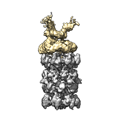 emd_0212.png emd_0212.png | 78.1 KB | ||
| Filedesc metadata | emd-0212.cif.gz | 6.2 KB | ||
| Others | emd_0212_additional.map.gz | 1.8 MB | ||
| Archive directory |  http://ftp.pdbj.org/pub/emdb/structures/EMD-0212ftp://ftp.pdbj.org/pub/emdb/structures/EMD-0212 http://ftp.pdbj.org/pub/emdb/structures/EMD-0212ftp://ftp.pdbj.org/pub/emdb/structures/EMD-0212 | HTTPS FTP |
-Related structure data
| Related structure data |  6he8MC  0209C  0210C 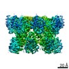 0211C 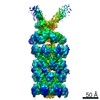 0213C 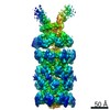 0214C 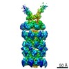 0215C 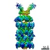 0216C 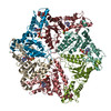 6he4C 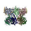 6he5C 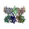 6he7C 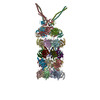 6he9C 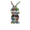 6heaC 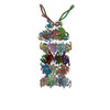 6hecC  6hedC M: atomic model generated by this map C: citing same article ( |
|---|---|
| Similar structure data |

-Links
| EMDB pages | EMDB (EBI/PDBe) / EMDataResource |
|---|---|
| Related items in Molecule of the Month |


-Map
| File | Download / File: emd_0212.map.gz / Format: CCP4 / Size: 216 MB / Type: IMAGE STORED AS FLOATING POINT NUMBER (4 BYTES) | ||||||||||||||||||||||||||||||||||||||||||||||||||||||||||||
|---|---|---|---|---|---|---|---|---|---|---|---|---|---|---|---|---|---|---|---|---|---|---|---|---|---|---|---|---|---|---|---|---|---|---|---|---|---|---|---|---|---|---|---|---|---|---|---|---|---|---|---|---|---|---|---|---|---|---|---|---|---|
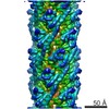
| Annotation | Pseudo-single capped PAN-proteasome in state 1 | ||||||||||||||||||||||||||||||||||||||||||||||||||||||||||||
| Projections & slices | Image control
Images are generated by Spider. | ||||||||||||||||||||||||||||||||||||||||||||||||||||||||||||
| Voxel size | X=Y=Z: 1.34 Å | ||||||||||||||||||||||||||||||||||||||||||||||||||||||||||||
| Density |
| ||||||||||||||||||||||||||||||||||||||||||||||||||||||||||||
| Symmetry | Space group: 1 | ||||||||||||||||||||||||||||||||||||||||||||||||||||||||||||
| Details | EMDB XML:
CCP4 map header:
| ||||||||||||||||||||||||||||||||||||||||||||||||||||||||||||
 Z (Sec.)
 Y (Row.)
 X (Col.)


-Supplemental data
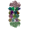
-Additional map: AAAob of PAN-proteasome in state 1
| File | emd_0212_additional.map | ||||||||||||
|---|---|---|---|---|---|---|---|---|---|---|---|---|---|
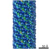
| Annotation | AAAob of PAN-proteasome in state 1 | ||||||||||||
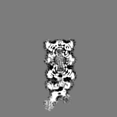
| Projections & Slices |
| ||||||||||||
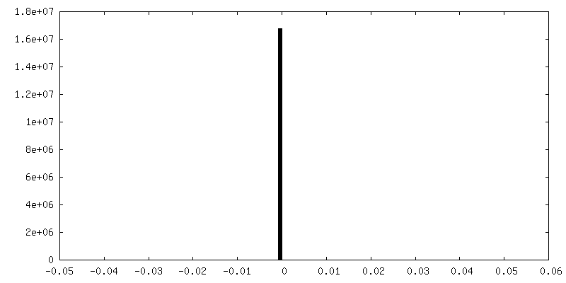
| Density Histograms |


- Sample components
-Entire : PAN-proteasome
| Entire | Name: PAN-proteasome |
|---|---|
| Components |
|
-Supramolecule #1: PAN-proteasome
| Supramolecule | Name: PAN-proteasome / type: complex / ID: 1 / Parent: 0 / Macromolecule list: #1-#3 |
|---|---|
| Source (natural) | Organism: Archaeoglobus fulgidus DSM 4304 (archaea) |
| Molecular weight | Theoretical: 1.25 MDa |
-Supramolecule #2: 20S-proteasome
| Supramolecule | Name: 20S-proteasome / type: complex / ID: 2 / Parent: 1 / Macromolecule list: #1-#2 |
|---|---|
| Source (natural) | Organism: Archaeoglobus fulgidus DSM 4304 (archaea) |
-Supramolecule #3: Proteasome-activating nucleotidase (PAN)
| Supramolecule | Name: Proteasome-activating nucleotidase (PAN) / type: complex / ID: 3 / Parent: 1 / Macromolecule list: #3 |
|---|---|
| Source (natural) | Organism: Archaeoglobus fulgidus DSM 4304 (archaea) |
-Macromolecule #1: Proteasome subunit alpha
| Macromolecule | Name: Proteasome subunit alpha / type: protein_or_peptide / ID: 1 / Number of copies: 14 / Enantiomer: LEVO / EC number: proteasome endopeptidase complex |
|---|---|
| Source (natural) | Organism: Archaeoglobus fulgidus (strain ATCC 49558 / VC-16 / DSM 4304 / JCM 9628 / NBRC 100126) (archaea) Strain: ATCC 49558 / VC-16 / DSM 4304 / JCM 9628 / NBRC 100126 |
| Molecular weight | Theoretical: 27.15616 KDa |
| Recombinant expression | Organism:  |
| Sequence | String: QMGYDRAITV FSPDGRLFQV EYAREAVKRG ATAIGIKCKE GVILIADKRV GSKLLEADTI EKIYKIDEHI CAATSGLVAD ARVLIDRAR IEAQINRLTY DEPITVKELA KKICDFKQQY TQYGGVRPFG VSLLIAGVDE VPKLYETDPS GALLEYKATA I GMGRNAVT ...String: QMGYDRAITV FSPDGRLFQV EYAREAVKRG ATAIGIKCKE GVILIADKRV GSKLLEADTI EKIYKIDEHI CAATSGLVAD ARVLIDRAR IEAQINRLTY DEPITVKELA KKICDFKQQY TQYGGVRPFG VSLLIAGVDE VPKLYETDPS GALLEYKATA I GMGRNAVT EFFEKEYRDD LSFDDAMVLG LVAMGLSIES ELVPENIEVG YVKVDDRTFK EVSPEELKPY VERANERIRE LL KK UniProtKB: Proteasome subunit alpha |
-Macromolecule #2: Proteasome subunit beta
| Macromolecule | Name: Proteasome subunit beta / type: protein_or_peptide / ID: 2 / Number of copies: 14 / Enantiomer: LEVO / EC number: proteasome endopeptidase complex |
|---|---|
| Source (natural) | Organism: Archaeoglobus fulgidus (strain ATCC 49558 / VC-16 / DSM 4304 / JCM 9628 / NBRC 100126) (archaea) Strain: ATCC 49558 / VC-16 / DSM 4304 / JCM 9628 / NBRC 100126 |
| Molecular weight | Theoretical: 22.132283 KDa |
| Recombinant expression | Organism: |
| Sequence | String: TTTVGLVCKD GVVMATEKRA TMGNFIASKA AKKIYQIADR MAMTTAGSVG DAQFLARIIK IEANLYEIRR ERKPTVRAIA TLTSNLLNS YRYFPYLVQL LIGGIDSEGK SIYSIDPIGG AIEEKDIVAT GSGSLTAYGV LEDRFTPEIG VDEAVELAVR A IYSAMKRD ...String: TTTVGLVCKD GVVMATEKRA TMGNFIASKA AKKIYQIADR MAMTTAGSVG DAQFLARIIK IEANLYEIRR ERKPTVRAIA TLTSNLLNS YRYFPYLVQL LIGGIDSEGK SIYSIDPIGG AIEEKDIVAT GSGSLTAYGV LEDRFTPEIG VDEAVELAVR A IYSAMKRD SASGDGIDVV KITEDEFYQY SPEEVEQILA KFRK UniProtKB: Proteasome subunit beta |
-Macromolecule #3: Proteasome-activating nucleotidase
| Macromolecule | Name: Proteasome-activating nucleotidase / type: protein_or_peptide / ID: 3 / Number of copies: 6 / Enantiomer: LEVO |
|---|---|
| Source (natural) | Organism: Archaeoglobus fulgidus (strain ATCC 49558 / VC-16 / DSM 4304 / JCM 9628 / NBRC 100126) (archaea) Strain: ATCC 49558 / VC-16 / DSM 4304 / JCM 9628 / NBRC 100126 |
| Molecular weight | Theoretical: 44.102977 KDa |
| Recombinant expression | Organism: |
| Sequence | String: LLEKLKKLEE DYYKLRELYR RLEDEKKFIE SERIRYEREV RRLRSEVERL RSPPLLVGVV SDILEDGRVV VKSSTGPKFV VNTSQYINE EELKPGARVA LNQQTLAIVN VLPTSKDPMV YGFEVEEKPE VSYEDIGGLD VQIEEIREAV ELPLLKPELF A EVGIEPPK ...String: LLEKLKKLEE DYYKLRELYR RLEDEKKFIE SERIRYEREV RRLRSEVERL RSPPLLVGVV SDILEDGRVV VKSSTGPKFV VNTSQYINE EELKPGARVA LNQQTLAIVN VLPTSKDPMV YGFEVEEKPE VSYEDIGGLD VQIEEIREAV ELPLLKPELF A EVGIEPPK GVLLYGPPGT GKTLLAKAVA NQTRATFIRV VGSEFVQKYI GEGARLVREV FQLAKEKAPS IIFIDELDAI AA RRTNSDT SGDREVQRTM MQLLAELDGF DPRGDVKVIG ATNRIDILDP AILRPGRFDR IIEVPLPTFE GRIQIFKIHT RKM KLAEDV DFKELARITE GASGADIKAI CTEAGMFAIR EERAKVTMLD FTKAIEKVLK KTTPIPDLKG VMFV UniProtKB: Proteasome-activating nucleotidase |
-Macromolecule #4: ADENOSINE-5'-TRIPHOSPHATE
| Macromolecule | Name: ADENOSINE-5'-TRIPHOSPHATE / type: ligand / ID: 4 / Number of copies: 4 / Formula: ATP |
|---|---|
| Molecular weight | Theoretical: 507.181 Da |
| Chemical component information |  ChemComp-ATP: |
-Macromolecule #5: MAGNESIUM ION
| Macromolecule | Name: MAGNESIUM ION / type: ligand / ID: 5 / Number of copies: 5 / Formula: MG |
|---|---|
| Molecular weight | Theoretical: 24.305 Da |
-Macromolecule #6: ADENOSINE-5'-DIPHOSPHATE
| Macromolecule | Name: ADENOSINE-5'-DIPHOSPHATE / type: ligand / ID: 6 / Number of copies: 1 / Formula: ADP |
|---|---|
| Molecular weight | Theoretical: 427.201 Da |
| Chemical component information |  ChemComp-ADP: |
-Experimental details
-Structure determination
| Method | cryo EM |
|---|---|
 Processing Processing | single particle reconstruction |
| Aggregation state | particle |
-Sample preparation
| Buffer | pH: 7.1 |
|---|---|
| Vitrification | Cryogen name: ETHANE-PROPANE |
- Electron microscopy
| Microscope | FEI TITAN KRIOS |
|---|---|
| Image recording | Film or detector model: GATAN K2 SUMMIT (4k x 4k) / Detector mode: SUPER-RESOLUTION / Average electron dose: 30.0 e/Å2 |
| Electron beam | Acceleration voltage: 300 kV / Electron source:  FIELD EMISSION GUN FIELD EMISSION GUN |
| Electron optics | Illumination mode: FLOOD BEAM / Imaging mode: BRIGHT FIELD |
| Experimental equipment |  Model: Titan Krios / Image courtesy: FEI Company |
-Image processing
| Startup model | Type of model: EMDB MAP |
|---|---|
| Final reconstruction | Applied symmetry - Point group: C1 (asymmetric) / Resolution.type: BY AUTHOR / Resolution: 6.86 Å / Resolution method: FSC 0.143 CUT-OFF / Software - Name: RELION (ver. 2.1) / Number images used: 38230 |
| Initial angle assignment | Type: PROJECTION MATCHING |
| Final angle assignment | Type: PROJECTION MATCHING |
-Atomic model buiding 1
| Refinement | Space: REAL / Protocol: FLEXIBLE FIT |
|---|---|
| Output model | PDB-6he8: |
