Movie
Movie Controller
 Structure viewers
 About Yorodumi Papers


+Search query
-Structure paper
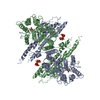
| Title | Structural mechanism underlying PHO1;H1-mediated phosphate transport in Arabidopsis. |
|---|---|
| Journal, issue, pages | Nat Plants, Vol. 11, Issue 2, Page 309-320, Year 2025 |
| Publish date | Jan 21, 2025 |
 Authors Authors | Sunzhenhe Fang / Yang Yang / Xue Zhang / Zhao Yang / Minhua Zhang / Yang Zhao / Chensi Zhang / Fang Yu / Yong-Fei Wang / Peng Zhang /  |
| PubMed Abstract | Arabidopsis PHOSPHATE 1 (AtPHO1) and its closest homologue AtPHO1;H1 are phosphate transporters that load phosphate into the xylem vessel for root-to-shoot translocation. AtPHO1 and AtPHO1;H1 are ...Arabidopsis PHOSPHATE 1 (AtPHO1) and its closest homologue AtPHO1;H1 are phosphate transporters that load phosphate into the xylem vessel for root-to-shoot translocation. AtPHO1 and AtPHO1;H1 are prototypical members of the unique SPX-EXS family, whose structural and molecular mechanisms remain elusive. In this study, we determined the cryogenic electron microscopy structure of AtPHO1;H1 binding with inorganic phosphate (Pi) and inositol hexakisphosphate in a closed conformation. Further molecular dynamic simulation and AlphaFold prediction support an open conformation. AtPHO1;H1 forms a domain-swapped homodimer that involves both the transmembrane ERD1/XPR1/SYG1 (EXS) domain and the cytoplasmic SYG1/Pho81/XPR1 (SPX) domain. The EXS domain presented by the SPX-EXS family represents a novel protein fold, and an independent substrate transport pathway and substrate-binding site are present in each EXS domain. Two gating residues, Trp719 and Tyr610, are identified above the substrate-binding site to control opening and closing of the pathway. The SPX domain features positively charged patches and/or residues at the dimer interface to accommodate inositol hexakisphosphate molecules, whose binding mediates dimerization and enhances AtPHO1;H1 activity. In addition, a C-terminal tail is required for AtPHO1;H1 activity. On the basis of structural and functional analysis, a working model for Pi efflux mediated by AtPHO1;H1 and its homologues was postulated, suggesting a channel-like mechanism. This study not only reveals the molecular and regulatory mechanism underlying Pi transport mediated by the unique SPX-EXS family, but also provides potential for crop engineering to enhance phosphorus-use efficiency in sustainable agriculture. |
 External links External links | Nat Plants / PubMed:39838070 |
| Methods | EM (single particle) |
| Resolution | 3.05 - 3.34 Å |
| Structure data | EMDB-60648, PDB-9ik4: EMDB-61430: Cryo-EM structure of the EXS domain of Arabidopsis thaliana phosphatetransporter PHO1;H1 |
| Chemicals |  ChemComp-IHP:  ChemComp-PO4: |
| Source |
|
 Keywords Keywords | MEMBRANE PROTEIN / phosphate transport / SPX domain / SPX-EXS / InsP6 / PHO1 / cryo-EM |