 Movie
Movie Controller

[English] 日本語
 Yorodumi
- EMDB-60648: Cryo-EM structure of Arabidopsis thaliana phosphate transporter P... -
+ Open data

- Basic information
| Entry | 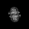 | |||||||||
|---|---|---|---|---|---|---|---|---|---|---|
| Title | Cryo-EM structure of Arabidopsis thaliana phosphate transporter PHO1;H1 | |||||||||
 Map data Map data | ||||||||||
 Sample Sample |
| |||||||||
 Keywords Keywords | phosphate transport / SPX domain / SPX-EXS / InsP6 / PHO1 / MEMBRANE PROTEIN | |||||||||
| Function / homology |  Function and homology information Function and homology informationphosphate transmembrane transporter activity / phosphate ion transport / cellular response to phosphate starvation / inositol hexakisphosphate binding / trans-Golgi network / plasma membrane Similarity search - Function | |||||||||
| Biological species |  | |||||||||
| Method | single particle reconstruction / cryo EM / Resolution: 3.34 Å | |||||||||
 Authors Authors | Fang S / Zhang X / Zhang P | |||||||||
| Funding support |  China, 1 items China, 1 items
| |||||||||
 Citation Citation | Journal: Nat Plants / Year: 2025 Title: Structural mechanism underlying PHO1;H1-mediated phosphate transport in Arabidopsis. Authors: Sunzhenhe Fang / Yang Yang / Xue Zhang / Zhao Yang / Minhua Zhang / Yang Zhao / Chensi Zhang / Fang Yu / Yong-Fei Wang / Peng Zhang / Abstract: Arabidopsis PHOSPHATE 1 (AtPHO1) and its closest homologue AtPHO1;H1 are phosphate transporters that load phosphate into the xylem vessel for root-to-shoot translocation. AtPHO1 and AtPHO1;H1 are ...Arabidopsis PHOSPHATE 1 (AtPHO1) and its closest homologue AtPHO1;H1 are phosphate transporters that load phosphate into the xylem vessel for root-to-shoot translocation. AtPHO1 and AtPHO1;H1 are prototypical members of the unique SPX-EXS family, whose structural and molecular mechanisms remain elusive. In this study, we determined the cryogenic electron microscopy structure of AtPHO1;H1 binding with inorganic phosphate (Pi) and inositol hexakisphosphate in a closed conformation. Further molecular dynamic simulation and AlphaFold prediction support an open conformation. AtPHO1;H1 forms a domain-swapped homodimer that involves both the transmembrane ERD1/XPR1/SYG1 (EXS) domain and the cytoplasmic SYG1/Pho81/XPR1 (SPX) domain. The EXS domain presented by the SPX-EXS family represents a novel protein fold, and an independent substrate transport pathway and substrate-binding site are present in each EXS domain. Two gating residues, Trp719 and Tyr610, are identified above the substrate-binding site to control opening and closing of the pathway. The SPX domain features positively charged patches and/or residues at the dimer interface to accommodate inositol hexakisphosphate molecules, whose binding mediates dimerization and enhances AtPHO1;H1 activity. In addition, a C-terminal tail is required for AtPHO1;H1 activity. On the basis of structural and functional analysis, a working model for Pi efflux mediated by AtPHO1;H1 and its homologues was postulated, suggesting a channel-like mechanism. This study not only reveals the molecular and regulatory mechanism underlying Pi transport mediated by the unique SPX-EXS family, but also provides potential for crop engineering to enhance phosphorus-use efficiency in sustainable agriculture. | |||||||||
| History |
|
- Structure visualization
| Supplemental images |
|---|

- Downloads & links
-EMDB archive
| Map data | emd_60648.map.gz | 106.8 MB | EMDB map data format | |
|---|---|---|---|---|
| Header (meta data) | emd-60648-v30.xmlemd-60648.xml | 19.6 KB 19.6 KB | Display Display | EMDB header |
| FSC (resolution estimation) | emd_60648_fsc.xml | 10.2 KB | Display | FSC data file |
| Images |  emd_60648.png emd_60648.png | 98.2 KB | ||
| Filedesc metadata | emd-60648.cif.gz | 6.4 KB | ||
| Others | emd_60648_additional_1.map.gzemd_60648_half_map_1.map.gzemd_60648_half_map_2.map.gz | 54.6 MB 105.1 MB 105.1 MB | ||
| Archive directory |  http://ftp.pdbj.org/pub/emdb/structures/EMD-60648ftp://ftp.pdbj.org/pub/emdb/structures/EMD-60648 http://ftp.pdbj.org/pub/emdb/structures/EMD-60648ftp://ftp.pdbj.org/pub/emdb/structures/EMD-60648 | HTTPS FTP |
-Related structure data
| Related structure data | 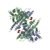 9ik4MC  9jf8C M: atomic model generated by this map C: citing same article ( |
|---|---|
| Similar structure data |
-Links
| EMDB pages | EMDB (EBI/PDBe) / EMDataResource |
|---|
-Map
| File | Download / File: emd_60648.map.gz / Format: CCP4 / Size: 113.6 MB / Type: IMAGE STORED AS FLOATING POINT NUMBER (4 BYTES) | ||||||||||||||||||||||||||||||||||||
|---|---|---|---|---|---|---|---|---|---|---|---|---|---|---|---|---|---|---|---|---|---|---|---|---|---|---|---|---|---|---|---|---|---|---|---|---|---|
| Projections & slices | Image control
Images are generated by Spider. | ||||||||||||||||||||||||||||||||||||
| Voxel size | X=Y=Z: 0.83 Å | ||||||||||||||||||||||||||||||||||||
| Density |
| ||||||||||||||||||||||||||||||||||||
| Symmetry | Space group: 1 | ||||||||||||||||||||||||||||||||||||
| Details | EMDB XML:
|
 Z (Sec.)
 Y (Row.)
 X (Col.)


-Supplemental data
-Additional map: unsharpen map
| File | emd_60648_additional_1.map | ||||||||||||
|---|---|---|---|---|---|---|---|---|---|---|---|---|---|
| Annotation | unsharpen map | ||||||||||||
| Projections & Slices |
| ||||||||||||
| Density Histograms |


-Half map: #2
| File | emd_60648_half_map_1.map | ||||||||||||
|---|---|---|---|---|---|---|---|---|---|---|---|---|---|
| Projections & Slices |
| ||||||||||||
| Density Histograms |


-Half map: #1
| File | emd_60648_half_map_2.map | ||||||||||||
|---|---|---|---|---|---|---|---|---|---|---|---|---|---|
| Projections & Slices |
| ||||||||||||
| Density Histograms |


- Sample components
-Entire : Phosphate transporter PHO1;H1
| Entire | Name: Phosphate transporter PHO1;H1 |
|---|---|
| Components |
|
-Supramolecule #1: Phosphate transporter PHO1;H1
| Supramolecule | Name: Phosphate transporter PHO1;H1 / type: complex / ID: 1 / Parent: 0 / Macromolecule list: #1 |
|---|---|
| Source (natural) | Organism: |
-Macromolecule #1: Phosphate transporter PHO1 homolog 1
| Macromolecule | Name: Phosphate transporter PHO1 homolog 1 / type: protein_or_peptide / ID: 1 / Number of copies: 2 / Enantiomer: LEVO |
|---|---|
| Source (natural) | Organism: |
| Molecular weight | Theoretical: 89.402562 KDa |
| Recombinant expression | Organism:   Spodoptera frugiperda (fall armyworm) Spodoptera frugiperda (fall armyworm) |
| Sequence | String: MVKFTKQFEG QLVPEWKDAF VDYSQLKKDL KKIHLFTNGV EKKHTETSLI KTVKSSLGRL SIFGNKGREQ SRVIQVHKKL ASSGSNNDV YETELLEKIA DDTDAAKEFF ACLDMQLNKV NQFYKTKEKE FLERGECLKK QMDILIELKD AFKQKQANGE S TQESKEDD ...String: MVKFTKQFEG QLVPEWKDAF VDYSQLKKDL KKIHLFTNGV EKKHTETSLI KTVKSSLGRL SIFGNKGREQ SRVIQVHKKL ASSGSNNDV YETELLEKIA DDTDAAKEFF ACLDMQLNKV NQFYKTKEKE FLERGECLKK QMDILIELKD AFKQKQANGE S TQESKEDD SISCTISCEY DSVRGRTEEM QLQVSCLDNL EDNGEEALES LGSEEPIKAN NEDSKLTTVS SRVFSCQGKN VK IKIPLTN PSRTFSAISY LINQSSSKKN GPDGGNKLQI SKKKLSHAEK MIKGALTELF KGLNYLKTYR NLNILAFMNI LKK FDKVTG KQILPIYLKV VESSYFNISD KVMILSDEVE EWFIKHLAGE NRRKAMKYLK PHHRKESHSV TFFIGLFTGC FVAL LAGYI IVAHLTGMYR QHSANTFYME TAYPVLSMFG LLFLHLFLYG CNIFMWRKAR INYSFIFELG SKNELKYRDV FLICT ASMS AIAGVMFVHL SLLEKGYSFR QVQVIPGLLL LGFLLILICP LNIFYKSSRY RLISVIRNIV FSPLYKVVML DFFMAD QLC SQVPMLRNLE YIACYYITGS YATQDYEYCM RVKYYRDLAY AVSFLPYYWR AMQCARRWFD EGETSHLVNL GKYVSAM LA AGTKVAYEKE RSLGWLCLVV AMSSVATIYQ LYWDFVKDWG LLQHNSNNPW LRNQLMLRQK SIYYFSMVLN LVLRLAWL Q TVLHSSFEHV DYRVTGLFLA ALEVIRRGQW NFYRLENEHL NNAGKFRAVK T UniProtKB: Phosphate transporter PHO1 homolog 1 |
-Macromolecule #2: INOSITOL HEXAKISPHOSPHATE
| Macromolecule | Name: INOSITOL HEXAKISPHOSPHATE / type: ligand / ID: 2 / Number of copies: 2 / Formula: IHP |
|---|---|
| Molecular weight | Theoretical: 660.035 Da |
| Chemical component information |  ChemComp-IHP: |
-Macromolecule #3: PHOSPHATE ION
| Macromolecule | Name: PHOSPHATE ION / type: ligand / ID: 3 / Number of copies: 2 / Formula: PO4 |
|---|---|
| Molecular weight | Theoretical: 94.971 Da |
| Chemical component information |  ChemComp-PO4: |
-Experimental details
-Structure determination
| Method | cryo EM |
|---|---|
 Processing Processing | single particle reconstruction |
| Aggregation state | particle |
-Sample preparation
| Buffer | pH: 7.5 |
|---|---|
| Vitrification | Cryogen name: ETHANE |
- Electron microscopy
| Microscope | FEI TITAN KRIOS |
|---|---|
| Image recording | Film or detector model: GATAN K3 BIOCONTINUUM (6k x 4k) / Average electron dose: 58.5 e/Å2 |
| Electron beam | Acceleration voltage: 300 kV / Electron source:  FIELD EMISSION GUN FIELD EMISSION GUN |
| Electron optics | Illumination mode: SPOT SCAN / Imaging mode: BRIGHT FIELD / Nominal defocus max: 20.0 µm / Nominal defocus min: 8.0 µm |
| Experimental equipment |  Model: Titan Krios / Image courtesy: FEI Company |

