Movie
Movie Controller
 Structure viewers
 About Yorodumi Papers


+Search query
-Structure paper
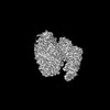
| Title | 2.6-Å resolution cryo-EM structure of a class Ia ribonucleotide reductase trapped with mechanism-based inhibitor NCDP. |
|---|---|
| Journal, issue, pages | Proc Natl Acad Sci U S A, Vol. 121, Issue 45, Page e2417157121, Year 2024 |
| Publish date | Nov 5, 2024 |
 Authors Authors | Dana E Westmoreland / Patricia R Feliciano / Gyunghoon Kang / Chang Cui / Albert Kim / JoAnne Stubbe / Daniel G Nocera / Catherine L Drennan /  |
| PubMed Abstract | Ribonucleotide reductases (RNRs) reduce ribonucleotides to deoxyribonucleotides using radical-based chemistry. For class Ia RNRs, the radical species is stored in a separate subunit (β2) from the ...Ribonucleotide reductases (RNRs) reduce ribonucleotides to deoxyribonucleotides using radical-based chemistry. For class Ia RNRs, the radical species is stored in a separate subunit (β2) from the subunit housing the active site (α2), requiring the formation of a short-lived α2β2 complex and long-range radical transfer (RT). RT occurs via proton-coupled electron transfer (PCET) over a long distance (~32-Å) and involves the formation and decay of multiple amino acid radical species. Here, we use cryogenic electron microscopy and a mechanism-based inhibitor 2'-azido-2'-deoxycytidine-5'-diphosphate (NCDP) to trap a wild-type α2β2 complex of class Ia RNR. We find that one α subunit has turned over and that the other is trapped, bound to β in a midturnover state. Instead of NCDP in the active site, forward RT has resulted in N loss, migration of the third nitrogen from the ribose C2' to C3' positions, and attachment of this nitrogen to the sulfur of cysteine-225. In this study, an inhibitor has been visualized as an adduct to an RNR. Additionally, this structure reveals the positions of PCET residues following forward RT, complementing the previous structure that depicted a preturnover PCET pathway and suggesting how PCET is gated at the α-β interface. This NCDP-trapped structure is also of sufficient resolution (2.6 Å) to visualize water molecules, allowing us to evaluate the proposal that water molecules are proton acceptors and donors as part of the PCET process. |
 External links External links | Proc Natl Acad Sci U S A / PubMed:39475643 / PubMed Central |
| Methods | EM (single particle) |
| Resolution | 2.6 Å |
| Structure data | EMDB-46711, PDB-9db2: |
| Chemicals |  ChemComp-DTP:  ChemComp-MG:  ChemComp-ATP: 
ChemComp-UNL:  PDB-1a3l:  ChemComp-FEO:  ChemComp-HOH: |
| Source |
|
 Keywords Keywords | OXIDOREDUCTASE / ribonucleotide reductase / class Ia / mechanistic inhibition |