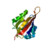
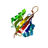
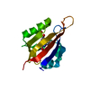
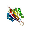
PDB-8qi8: Cryogenic temperature dark state structure of CrPhotLOV1 Method: X-RAY DIFFRACTION / Resolution: 1.35 Å
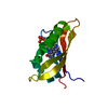
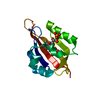
PDB-8qi9: CrPhotLOV1 dark state structure determined by serial synchrotron crystallography at room temperature Method: X-RAY DIFFRACTION / Resolution: 1.87 Å
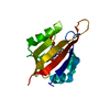
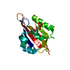
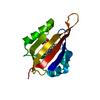
PDB-8qia: CrPhotLOV1 light state structure 2.5 ms (0-5 ms) after illumination determined by time-resolved serial synchrotron crystallography at room temperature Method: X-RAY DIFFRACTION / Resolution: 2.5 Å
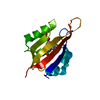
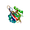
PDB-8qib: CrPhotLOV1 light state structure 7.5 ms (5-10 ms) after illumination determined by time-resolved serial synchrotron crystallography at room temperature Method: X-RAY DIFFRACTION / Resolution: 2.45 Å
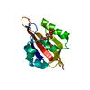
PDB-8qif: CrPhotLOV1 light state structure 12.5 ms (10-15 ms) after illumination determined by time-resolved serial synchrotron crystallography at room temperature Method: X-RAY DIFFRACTION / Resolution: 2.45 Å
PDB-8qig: CrPhotLOV1 light state structure 17.5 ms (15-20 ms) after illumination determined by time-resolved serial synchrotron crystallography at room temperature Method: X-RAY DIFFRACTION / Resolution: 2.5 Å
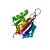
PDB-8qih: CrPhotLOV1 light state structure 22.5 ms (20-25 ms) after illumination determined by time-resolved serial synchrotron crystallography at room temperature Method: X-RAY DIFFRACTION / Resolution: 2.5 Å
PDB-8qii: CrPhotLOV1 light state structure 27.5 ms (25-30 ms) after illumination determined by time-resolved serial synchrotron crystallography at room temperature Method: X-RAY DIFFRACTION / Resolution: 2.5 Å
PDB-8qik: CrPhotLOV1 light state structure 32.5 ms (30-35 ms) after illumination determined by time-resolved serial synchrotron crystallography at room temperature Method: X-RAY DIFFRACTION / Resolution: 2.55 Å
PDB-8qil: CrPhotLOV1 light state structure 37.5 ms (35-40 ms) after illumination determined by time-resolved serial synchrotron crystallography at room temperature Method: X-RAY DIFFRACTION / Resolution: 2.55 Å
PDB-8qim: CrPhotLOV1 light state structure 42.5 ms (40-45 ms) after illumination determined by time-resolved serial synchrotron crystallography at room temperature Method: X-RAY DIFFRACTION / Resolution: 2.6 Å
PDB-8qin: CrPhotLOV1 light state structure 47.5 ms (45-50 ms) after illumination determined by time-resolved serial synchrotron crystallography at room temperature Method: X-RAY DIFFRACTION / Resolution: 2.7 Å
PDB-8qio: CrPhotLOV1 light state structure 52.5 ms (50-55 ms) after illumination determined by time-resolved serial synchrotron crystallography at room temperature Method: X-RAY DIFFRACTION / Resolution: 2.75 Å
PDB-8qip: CrPhotLOV1 light state structure 57.5 ms (55-60 ms) after illumination determined by time-resolved serial synchrotron crystallography at room temperature Method: X-RAY DIFFRACTION / Resolution: 2.7 Å
PDB-8qiq: CrPhotLOV1 light state structure 62.5 ms (60-65 ms) after illumination determined by time-resolved serial synchrotron crystallography at room temperature Method: X-RAY DIFFRACTION / Resolution: 2.9 Å
PDB-8qir: CrPhotLOV1 light state structure 67.5 ms (65-70 ms) after illumination determined by time-resolved serial synchrotron crystallography at room temperature Method: X-RAY DIFFRACTION / Resolution: 3.0 Å
PDB-8qis: CrPhotLOV1 light state structure 72.5 ms (70-75 ms) after illumination determined by time-resolved serial synchrotron crystallography at room temperature Method: X-RAY DIFFRACTION / Resolution: 2.9 Å
PDB-8qit: CrPhotLOV1 light state structure 77.5 ms (75-80 ms) after illumination determined by time-resolved serial synchrotron crystallography at room temperature Method: X-RAY DIFFRACTION / Resolution: 2.9 Å
PDB-8qiu: CrPhotLOV1 light state structure 82.5 ms (80-85 ms) after illumination determined by time-resolved serial synchrotron crystallography at room temperature Method: X-RAY DIFFRACTION / Resolution: 3.0 Å
PDB-8qiv: CrPhotLOV1 light state structure 87.5 ms (85-90 ms) after illumination determined by time-resolved serial synchrotron crystallography at room temperature Method: X-RAY DIFFRACTION / Resolution: 3.1 Å
PDB-8qiw: CrPhotLOV1 light state structure 92.5 ms (90-95 ms) after illumination determined by time-resolved serial synchrotron crystallography at room temperature Method: X-RAY DIFFRACTION / Resolution: 3.05 Å
In the structure databanks used in Yorodumi, some data are registered as the other names, "COVID-19 virus" and "2019-nCoV". Here are the details of the virus and the list of structure data.
Jan 31, 2019. EMDB accession codes are about to change! (news from PDBe EMDB page)
EMDB accession codes are about to change! (news from PDBe EMDB page)
The allocation of 4 digits for EMDB accession codes will soon come to an end. Whilst these codes will remain in use, new EMDB accession codes will include an additional digit and will expand incrementally as the available range of codes is exhausted. The current 4-digit format prefixed with “EMD-” (i.e. EMD-XXXX) will advance to a 5-digit format (i.e. EMD-XXXXX), and so on. It is currently estimated that the 4-digit codes will be depleted around Spring 2019, at which point the 5-digit format will come into force.
The EM Navigator/Yorodumi systems omit the EMD- prefix.
Related info.:Q: What is EMD? / ID/Accession-code notation in Yorodumi/EM Navigator
 Movie
Movie Controller
 Structure viewers
 About Yorodumi Papers


 Authors
 External links
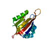


 Keywords
 chlamydomonas reinhardtii (plant)