 Movie
Movie Controller

[English] 日本語
 Yorodumi
- PDB-8qi9: CrPhotLOV1 dark state structure determined by serial synchrotron ... -
+ Open data

- Basic information
| Entry | Database: PDB / ID: 8qi9 | |||||||||||||||
|---|---|---|---|---|---|---|---|---|---|---|---|---|---|---|---|---|
| Title | CrPhotLOV1 dark state structure determined by serial synchrotron crystallography at room temperature | |||||||||||||||
 Components Components | Phototropin | |||||||||||||||
 Keywords Keywords | FLAVOPROTEIN / PHOTOTROPIN / FLAVIN / ELECTRON TRANSPORT | |||||||||||||||
| Function / homology |  Function and homology information Function and homology informationblue light signaling pathway / blue light photoreceptor activity / protein autophosphorylation / protein phosphorylation / non-specific serine/threonine protein kinase / protein serine kinase activity / protein serine/threonine kinase activity / ATP binding / nucleus / plasma membrane / cytoplasm Similarity search - Function | |||||||||||||||
| Biological species |   Chlamydomonas reinhardtii (plant) Chlamydomonas reinhardtii (plant) | |||||||||||||||
| Method |  X-RAY DIFFRACTION / SYNCHROTRON / MOLECULAR REPLACEMENT / Resolution: 1.87 Å X-RAY DIFFRACTION / SYNCHROTRON / MOLECULAR REPLACEMENT / Resolution: 1.87 Å | |||||||||||||||
 Authors Authors | Gotthard, G. / Mous, S. / Weinert, T. / Maia, R.N.A. / James, D. / Dworkowski, F. / Gashi, D. / Antonia, F. / Wang, M. / Panepucci, E. ...Gotthard, G. / Mous, S. / Weinert, T. / Maia, R.N.A. / James, D. / Dworkowski, F. / Gashi, D. / Antonia, F. / Wang, M. / Panepucci, E. / Ozerov, D. / Schertler, G.F.X. / Heberle, J. / Standfuss, J. / Nogly, P. | |||||||||||||||
| Funding support | European Union,  Switzerland, Switzerland,  Poland, 4items Poland, 4items
| |||||||||||||||
 Citation Citation | Journal: Iucrj / Year: 2024 Title: Capturing the blue-light activated state of the Phot-LOV1 domain from Chlamydomonas reinhardtii using time-resolved serial synchrotron crystallography. Authors: Gotthard, G. / Mous, S. / Weinert, T. / Maia, R.N.A. / James, D. / Dworkowski, F. / Gashi, D. / Furrer, A. / Ozerov, D. / Panepucci, E. / Wang, M. / Schertler, G.F.X. / Heberle, J. / Standfuss, J. / Nogly, P. | |||||||||||||||
| History |
|
- Structure visualization
| Structure viewer | Molecule: MolmilJmol/JSmol |
|---|
- Downloads & links
-Download
| PDBx/mmCIF format | 8qi9.cif.gz | 73.1 KB | Display | PDBx/mmCIF format |
|---|---|---|---|---|
| PDB format | pdb8qi9.ent.gz | 44.2 KB | Display | PDB format |
| PDBx/mmJSON format | 8qi9.json.gz | Tree view | PDBx/mmJSON format | |
| Others |  Other downloads Other downloads |
-Validation report
| Arichive directory | https://data.pdbj.org/pub/pdb/validation_reports/qi/8qi9ftp://data.pdbj.org/pub/pdb/validation_reports/qi/8qi9 | HTTPS FTP |
|---|
-Related structure data
| Related structure data | 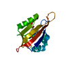 8qi8C  8qiaC 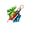 8qibC 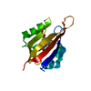 8qifC  8qigC 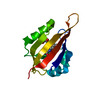 8qihC 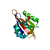 8qiiC 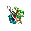 8qikC 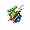 8qilC 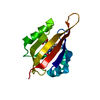 8qimC  8qinC 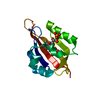 8qioC 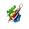 8qipC 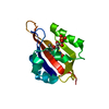 8qiqC  8qirC  8qisC  8qitC 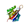 8qiuC  8qivC 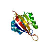 8qiwC C: citing same article ( |
|---|---|
| Similar structure data |
-Links
 PDBj
PDBj


- Assembly
| Deposited unit | 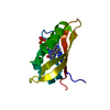
| ||||||||||||
|---|---|---|---|---|---|---|---|---|---|---|---|---|---|
| 1 |
| ||||||||||||
| 2 | 
| ||||||||||||
| Unit cell |
| ||||||||||||
| Components on special symmetry positions |
|
-Components
| #1: Protein | Mass: 15338.354 Da / Num. of mol.: 1 Source method: isolated from a genetically manipulated source Source: (gene. exp.) Chlamydomonas reinhardtii (plant) / Gene: PHOT, CHLRE_03g199000v5, CHLREDRAFT_183965 / Production host:  References: UniProt: Q8LPD9, non-specific serine/threonine protein kinase |
|---|---|
| #2: Chemical | ChemComp-FMN /   Mass: 456.344 Da / Num. of mol.: 1 / Source method: obtained synthetically / Formula: C17H21N4O9P / Feature type: SUBJECT OF INVESTIGATION Mass: 456.344 Da / Num. of mol.: 1 / Source method: obtained synthetically / Formula: C17H21N4O9P / Feature type: SUBJECT OF INVESTIGATION |
| #3: Water | ChemComp-HOH /  Mass: 18.015 Da / Num. of mol.: 102 / Source method: isolated from a natural source / Formula: H2O Mass: 18.015 Da / Num. of mol.: 102 / Source method: isolated from a natural source / Formula: H2O |
| Has ligand of interest | Y |
-Experimental details
-Experiment
| Experiment | Method: X-RAY DIFFRACTION / Number of used crystals: 1 |
|---|
- Sample preparation
| Crystal | Density Matthews: 4.11 Å3/Da / Density % sol: 70.04 % |
|---|---|
| Crystal grow | Temperature: 293 K / Method: batch mode / pH: 6.5 Details: 100 mM sodium cacodylate at pH 6.5 and 1.0 M sodium citrate dibasic trihydrate |
-Data collection
| Diffraction | Mean temperature: 293 K / Serial crystal experiment: N |
|---|---|
| Diffraction source | Source: SYNCHROTRON / Site: SLS / Beamline: X06SA / Wavelength: 1 Å |
| Detector | Type: DECTRIS EIGER X 16M / Detector: PIXEL / Date: Oct 16, 2019 |
| Radiation | Protocol: SINGLE WAVELENGTH / Monochromatic (M) / Laue (L): M / Scattering type: x-ray |
| Radiation wavelength | Wavelength: 1 Å / Relative weight: 1 |
| Reflection | Resolution: 1.87→104.7 Å / Num. obs: 17122 / % possible obs: 99.87 % / Redundancy: 1959.27 % / Biso Wilson estimate: 32.45 Å2 / CC1/2: 1 / CC star: 1 / R split: 0.0584 / Net I/σ(I): 13.25 |
| Reflection shell | Resolution: 1.87→1.9 Å / Redundancy: 310.4 % / Mean I/σ(I) obs: 0.73 / Num. unique obs: 1673 / CC1/2: 0.33 / CC star: 0.7 / R split: 1.3435 / % possible all: 99.1 |
- Processing
| Software |
| |||||||||||||||||||||||||||||||||||||||||||||||||
|---|---|---|---|---|---|---|---|---|---|---|---|---|---|---|---|---|---|---|---|---|---|---|---|---|---|---|---|---|---|---|---|---|---|---|---|---|---|---|---|---|---|---|---|---|---|---|---|---|---|---|
| Refinement | Method to determine structure: MOLECULAR REPLACEMENT / Resolution: 1.87→60.77 Å / SU ML: 0.2011 / Cross valid method: FREE R-VALUE / σ(F): 1.34 / Phase error: 21.9646 Stereochemistry target values: GeoStd + Monomer Library + CDL v1.2
| |||||||||||||||||||||||||||||||||||||||||||||||||
| Solvent computation | Shrinkage radii: 0.9 Å / VDW probe radii: 1.11 Å / Solvent model: FLAT BULK SOLVENT MODEL | |||||||||||||||||||||||||||||||||||||||||||||||||
| Displacement parameters | Biso mean: 40.14 Å2 | |||||||||||||||||||||||||||||||||||||||||||||||||
| Refinement step | Cycle: LAST / Resolution: 1.87→60.77 Å
| |||||||||||||||||||||||||||||||||||||||||||||||||
| Refine LS restraints |
| |||||||||||||||||||||||||||||||||||||||||||||||||
| LS refinement shell |
|
