[English] 日本語
Yorodumi Papers
- Database of articles cited by EMDB/PDB/SASBDB data -

+
Search query

Keywords
Structure methods
Author
Journal
IF

-
Structure paper

TitleStructure of the decoy module of human glycoprotein 2 and uromodulin and its interaction with bacterial adhesin FimH.
Journal, issue, pagesNat Struct Mol Biol, Vol. 29, Issue 3, Page 190-193, Year 2022
Publish dateMar 10, 2022
AuthorsAlena Stsiapanava / Chenrui Xu / Shunsuke Nishio / Ling Han / Nao Yamakawa / Marta Carroni / Kathryn Tunyasuvunakool / John Jumper / Daniele de Sanctis / Bin Wu / Luca Jovine /
PubMed AbstractGlycoprotein 2 (GP2) and uromodulin (UMOD) filaments protect against gastrointestinal and urinary tract infections by acting as decoys for bacterial fimbrial lectin FimH. By combining AlphaFold2 ...Glycoprotein 2 (GP2) and uromodulin (UMOD) filaments protect against gastrointestinal and urinary tract infections by acting as decoys for bacterial fimbrial lectin FimH. By combining AlphaFold2 predictions with X-ray crystallography and cryo-EM, we show that these proteins contain a bipartite decoy module whose new fold presents the high-mannose glycan recognized by FimH. The structure rationalizes UMOD mutations associated with kidney diseases and visualizes a key epitope implicated in cast nephropathy.
External linksNat Struct Mol Biol / PubMed:35273390 / PubMed Central
MethodsEM (single particle) / X-ray diffraction
Resolution1.35 - 7.4 Å
Structure data

EMDB-13378, PDB-7pfp:
Full-length cryo-EM structure of the native human uromodulin (UMOD)/Tamm-Horsfall protein (THP) filament
Method: EM (single particle) / Resolution: 6.1 Å

EMDB-13794, PDB-7q3n:
Cryo-EM of the complex between human uromodulin (UMOD)/Tamm-Horsfall protein (THP) and the FimH lectin domain from uropathogenic E. coli
Method: EM (single particle) / Resolution: 7.4 Å

PDB-7p6r:
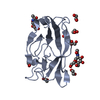
Crystal structure of the FimH-binding decoy module of human glycoprotein 2 (GP2) (crystal form I)
Method: X-RAY DIFFRACTION / Resolution: 1.9 Å

PDB-7p6s:
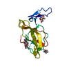
Crystal structure of the FimH-binding decoy module of human glycoprotein 2 (GP2) (crystal form II)
Method: X-RAY DIFFRACTION / Resolution: 1.35 Å

PDB-7p6t:
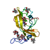
Crystal structure of the FimH-binding decoy module of human glycoprotein 2 (GP2) (crystal form III)
Method: X-RAY DIFFRACTION / Resolution: 1.4 Å

Chemicals

ChemComp-NAG:
2-acetamido-2-deoxy-beta-D-glucopyranose

ChemComp-EDO:
1,2-ETHANEDIOL

ChemComp-HOH:
WATER

ChemComp-9JE:
pentane-1,5-diol

ChemComp-9D2:
2-ethyl-2-(hydroxymethyl)propane-1,3-diol

Source
  • homo sapiens (human)
  • escherichia coli (strain uti89 / upec) (bacteria)
KeywordsANTIMICROBIAL PROTEIN / Extracellular matrix / glycoprotein / N-glycan / structural protein / decoy module / D10C domain / D8C sequence / pancreas / intestine / mucosal immunity / FimH / AlphaFold / EGF DOMAIN / BETA-HAIRPIN / ZP MODULE / ZP DOMAIN / ZP-N DOMAIN / ZP-C DOMAIN / INTERDOMAIN LINKER / PROTEIN FILAMENT / PROTEIN POLYMERIZATION / D8C DOMAIN / HIGH-MANNOSE SUGAR / CELL ADHESION / BACTERIAL ADHESIN / TYPE I PILUS / SUGAR BINDING PROTEIN / LECTIN / URINARY TRACT INFECTION / UTI / UROPATHOGENIC E. COLI / UPEC

+
About Yorodumi Papers

-
News

-
Feb 9, 2022. New format data for meta-information of EMDB entries

New format data for meta-information of EMDB entries

  • Version 3 of the EMDB header file is now the official format.
  • The previous official version 1.9 will be removed from the archive.

Related info.:EMDB header

External links:wwPDB to switch to version 3 of the EMDB data model

-
Aug 12, 2020. Covid-19 info

Covid-19 info

URL: https://pdbj.org/emnavi/covid19.php

New page: Covid-19 featured information page in EM Navigator.

Related info.:Covid-19 info / Mar 5, 2020. Novel coronavirus structure data

+
Mar 5, 2020. Novel coronavirus structure data

Novel coronavirus structure data

Related info.:Yorodumi Speices / Aug 12, 2020. Covid-19 info

External links:COVID-19 featured content - PDBj / Molecule of the Month (242):Coronavirus Proteases

+
Jan 31, 2019. EMDB accession codes are about to change! (news from PDBe EMDB page)

EMDB accession codes are about to change! (news from PDBe EMDB page)

  • The allocation of 4 digits for EMDB accession codes will soon come to an end. Whilst these codes will remain in use, new EMDB accession codes will include an additional digit and will expand incrementally as the available range of codes is exhausted. The current 4-digit format prefixed with “EMD-” (i.e. EMD-XXXX) will advance to a 5-digit format (i.e. EMD-XXXXX), and so on. It is currently estimated that the 4-digit codes will be depleted around Spring 2019, at which point the 5-digit format will come into force.
  • The EM Navigator/Yorodumi systems omit the EMD- prefix.

Related info.:Q: What is EMD? / ID/Accession-code notation in Yorodumi/EM Navigator

External links:EMDB Accession Codes are Changing Soon! / Contact to PDBj

+
Jul 12, 2017. Major update of PDB

Major update of PDB

  • wwPDB released updated PDB data conforming to the new PDBx/mmCIF dictionary.
  • This is a major update changing the version number from 4 to 5, and with Remediation, in which all the entries are updated.
  • In this update, many items about electron microscopy experimental information are reorganized (e.g. em_software).
  • Now, EM Navigator and Yorodumi are based on the updated data.

External links:wwPDB Remediation / Enriched Model Files Conforming to OneDep Data Standards Now Available in the PDB FTP Archive

-
Yorodumi Papers

Database of articles cited by EMDB/PDB/SASBDB data

  • Database of articles cited by EMDB, PDB, and SASBDB entries
  • Using PubMed data

Related info.:EMDB / PDB / SASBDB / Yorodumi / EMN Papers / Changes in new EM Navigator and Yorodumi

Read more