 Movie
Movie Controller
 Structure viewers
 About Yorodumi Papers


+Search query
-Structure paper
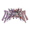
| Title | Structure and mechanism of human diacylglycerol O-acyltransferase 1. |
|---|---|
| Journal, issue, pages | Nature, Vol. 581, Issue 7808, Page 329-332, Year 2020 |
| Publish date | May 13, 2020 |
 Authors Authors | Lie Wang / Hongwu Qian / Yin Nian / Yimo Han / Zhenning Ren / Hanzhi Zhang / Liya Hu / B V Venkataram Prasad / Arthur Laganowsky / Nieng Yan / Ming Zhou /   |
| PubMed Abstract | Diacylglycerol O-acyltransferase 1 (DGAT1) synthesizes triacylglycerides and is required for dietary fat absorption and fat storage in humans. DGAT1 belongs to the membrane-bound O-acyltransferase ...Diacylglycerol O-acyltransferase 1 (DGAT1) synthesizes triacylglycerides and is required for dietary fat absorption and fat storage in humans. DGAT1 belongs to the membrane-bound O-acyltransferase (MBOAT) superfamily, members of which are found in all kingdoms of life and are involved in the acylation of lipids and proteins. How human DGAT1 and other mammalian members of the MBOAT family recognize their substrates and catalyse their reactions is unknown. The absence of three-dimensional structures also hampers rational targeting of DGAT1 for therapeutic purposes. Here we present the cryo-electron microscopy structure of human DGAT1 in complex with an oleoyl-CoA substrate. Each DGAT1 protomer has nine transmembrane helices, eight of which form a conserved structural fold that we name the MBOAT fold. The MBOAT fold in DGAT1 forms a hollow chamber in the membrane that encloses highly conserved catalytic residues. The chamber has separate entrances for each of the two substrates, fatty acyl-CoA and diacylglycerol. DGAT1 can exist as either a homodimer or a homotetramer and the two forms have similar enzymatic activity. The N terminus of DGAT1 interacts with the neighbouring protomer and these interactions are required for enzymatic activity. |
 External links External links | Nature / PubMed:32433610 / PubMed Central |
| Methods | EM (single particle) |
| Resolution | 3.1 Å |
| Structure data | EMDB-21302, PDB-6vp0: |
| Chemicals |  ChemComp-POV:  ChemComp-P5S:  ChemComp-3VV:  ChemComp-AV0: |
| Source |
|
 Keywords Keywords | MEMBRANE PROTEIN / diacylglycerol acyltransferase 1 / MBOAT / oleoyl-CoA / ER |

 homo sapiens (human)