 Movie
Movie Controller
 Structure viewers
 About Yorodumi Papers


+Search query
-Structure paper
| Title | Nanobeam precession-assisted 3D electron diffraction reveals a new polymorph of hen egg-white lysozyme. |
|---|---|
| Journal, issue, pages | IUCrJ, Vol. 6, Issue Pt 2, Page 178-188, Year 2019 |
| Publish date | Mar 1, 2019 |
 Authors Authors | Arianna Lanza / Eleonora Margheritis / Enrico Mugnaioli / Valentina Cappello / Gianpiero Garau / Mauro Gemmi /  |
| PubMed Abstract | Recent advances in 3D electron diffraction have allowed the structure determination of several model proteins from submicrometric crystals, the unit-cell parameters and structures of which could be ...Recent advances in 3D electron diffraction have allowed the structure determination of several model proteins from submicrometric crystals, the unit-cell parameters and structures of which could be immediately validated by known models previously obtained by X-ray crystallography. Here, the first new protein structure determined by 3D electron diffraction data is presented: a previously unobserved polymorph of hen egg-white lysozyme. This form, with unit-cell parameters = 31.9, = 54.4, = 71.8 Å, β = 98.8°, grows as needle-shaped submicrometric crystals simply by vapor diffusion starting from previously reported crystallization conditions. Remarkably, the data were collected using a low-dose stepwise experimental setup consisting of a precession-assisted nanobeam of ∼150 nm, which has never previously been applied for solving protein structures. The crystal structure was additionally validated using X-ray synchrotron-radiation sources by both powder diffraction and single-crystal micro-diffraction. 3D electron diffraction can be used for the structural characterization of submicrometric macromolecular crystals and is able to identify novel protein polymorphs that are hardly visible in conventional X-ray diffraction experiments. Additionally, the analysis, which was performed on both nanocrystals and microcrystals from the same crystallization drop, suggests that an integrated view from 3D electron diffraction and X-ray microfocus diffraction can be applied to obtain insights into the molecular dynamics during protein crystal growth. |
 External links External links | IUCrJ / PubMed:30867915 / PubMed Central |
| Methods | X-ray diffraction / EM (electron crystallography) |
| Resolution | 2.6 - 2.8 Å |
| Structure data | 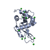 PDB-6ht2: 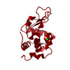 PDB-6hu5: |
| Chemicals |  ChemComp-CL:  ChemComp-HOH: |
| Source |
|
 Keywords Keywords | HYDROLASE / MICROFOCUS / LYSOZYME / HEWL / ED / ELECTRON / DIFFRACTION / CRYSTAL / CHLORIDE / ---- / HALOGEN / PROTEIN / DIMER |

