[English] 日本語
Yorodumi Papers
- Database of articles cited by EMDB/PDB/SASBDB data -

+
Search query

Keywords
Structure methods
Author
Journal
IF

-
Structure paper

TitleDead-end complex, lipid interactions and catalytic mechanism of microsomal glutathione transferase 1, an electron crystallography and mutagenesis investigation.
Journal, issue, pagesSci Rep, Vol. 7, Issue 1, Page 7897, Year 2017
Publish dateAug 11, 2017
AuthorsQie Kuang / Pasi Purhonen / Johan Ålander / Richard Svensson / Veronika Hoogland / Jens Winerdal / Linda Spahiu / Astrid Ottosson-Wadlund / Caroline Jegerschöld / Ralf Morgenstern / Hans Hebert /
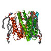
PubMed AbstractMicrosomal glutathione transferase 1 (MGST1) is a detoxification enzyme belonging to the Membrane Associated Proteins in Eicosanoid and Glutathione Metabolism (MAPEG) superfamily. Here we have used ...Microsomal glutathione transferase 1 (MGST1) is a detoxification enzyme belonging to the Membrane Associated Proteins in Eicosanoid and Glutathione Metabolism (MAPEG) superfamily. Here we have used electron crystallography of two-dimensional crystals in order to determine an atomic model of rat MGST1 in a lipid environment. The model comprises 123 of the 155 amino acid residues, two structured phospholipid molecules, two aliphatic chains and one glutathione (GSH) molecule. The functional unit is a homotrimer centered on the crystallographic three-fold axes of the unit cell. The GSH substrate binds in an extended conformation at the interface between two subunits of the trimer supported by new in vitro mutagenesis data. Mutation of Arginine 130 to alanine resulted in complete loss of activity consistent with a role for Arginine 130 in stabilizing the strongly nucleophilic GSH thiolate required for catalysis. Based on the new model and an electron diffraction data set from crystals soaked with trinitrobenzene, that forms a dead-end Meisenheimer complex with GSH, a difference map was calculated. The map reveals side chain movements opening a cavity that defines the second substrate site.
External linksSci Rep / PubMed:28801553 / PubMed Central
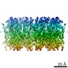
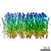
MethodsEM (electron crystallography)
Resolution3.5 Å
Structure data

EMDB-8076, PDB-5i9k:
The structure of microsomal glutathione transferase 1
Method: EM (electron crystallography) / Resolution: 3.5 Å

EMDB-8084, PDB-5ia9:
The structure of microsomal glutathione transferase 1 in complex with Meisenheimer complex
Method: EM (electron crystallography) / Resolution: 3.5 Å

Chemicals

ChemComp-GSH:
GLUTATHIONE / Glutathione

ChemComp-PC1:
1,2-DIACYL-SN-GLYCERO-3-PHOSPHOCHOLINE / phospholipid*YM / Phosphatidylcholine

ChemComp-PLM:
PALMITIC ACID / Palmitic acid

ChemComp-GTD:
1-(S-GLUTATHIONYL)-2,4,6-TRINITROCYCLOHEXA-2,5-DIENE

Source
  • rattus norvegicus (Norway rat)
KeywordsTRANSFERASE / Membrane / Enzyme / Four-helical bundle / Trimer / Meisenheimer complex

+
About Yorodumi Papers

-
News

-
Feb 9, 2022. New format data for meta-information of EMDB entries

New format data for meta-information of EMDB entries

  • Version 3 of the EMDB header file is now the official format.
  • The previous official version 1.9 will be removed from the archive.

Related info.:EMDB header

External links:wwPDB to switch to version 3 of the EMDB data model

-
Aug 12, 2020. Covid-19 info

Covid-19 info

URL: https://pdbj.org/emnavi/covid19.php

New page: Covid-19 featured information page in EM Navigator.

Related info.:Covid-19 info / Mar 5, 2020. Novel coronavirus structure data

+
Mar 5, 2020. Novel coronavirus structure data

Novel coronavirus structure data

Related info.:Yorodumi Speices / Aug 12, 2020. Covid-19 info

External links:COVID-19 featured content - PDBj / Molecule of the Month (242):Coronavirus Proteases

+
Jan 31, 2019. EMDB accession codes are about to change! (news from PDBe EMDB page)

EMDB accession codes are about to change! (news from PDBe EMDB page)

  • The allocation of 4 digits for EMDB accession codes will soon come to an end. Whilst these codes will remain in use, new EMDB accession codes will include an additional digit and will expand incrementally as the available range of codes is exhausted. The current 4-digit format prefixed with “EMD-” (i.e. EMD-XXXX) will advance to a 5-digit format (i.e. EMD-XXXXX), and so on. It is currently estimated that the 4-digit codes will be depleted around Spring 2019, at which point the 5-digit format will come into force.
  • The EM Navigator/Yorodumi systems omit the EMD- prefix.

Related info.:Q: What is EMD? / ID/Accession-code notation in Yorodumi/EM Navigator

External links:EMDB Accession Codes are Changing Soon! / Contact to PDBj

+
Jul 12, 2017. Major update of PDB

Major update of PDB

  • wwPDB released updated PDB data conforming to the new PDBx/mmCIF dictionary.
  • This is a major update changing the version number from 4 to 5, and with Remediation, in which all the entries are updated.
  • In this update, many items about electron microscopy experimental information are reorganized (e.g. em_software).
  • Now, EM Navigator and Yorodumi are based on the updated data.

External links:wwPDB Remediation / Enriched Model Files Conforming to OneDep Data Standards Now Available in the PDB FTP Archive

-
Yorodumi Papers

Database of articles cited by EMDB/PDB/SASBDB data

  • Database of articles cited by EMDB, PDB, and SASBDB entries
  • Using PubMed data

Related info.:EMDB / PDB / SASBDB / Yorodumi / EMN Papers / Changes in new EM Navigator and Yorodumi

Read more