 Movie
Movie Controller
 Structure viewers
 About Yorodumi Papers


+Search query
-Structure paper
| Title | Conformational changes during pore formation by the perforin-related protein pleurotolysin. |
|---|---|
| Journal, issue, pages | PLoS Biol, Vol. 13, Issue 2, Page e1002049, Year 2015 |
| Publish date | Feb 5, 2015 |
 Authors Authors | Natalya Lukoyanova / Stephanie C Kondos / Irene Farabella / Ruby H P Law / Cyril F Reboul / Tom T Caradoc-Davies / Bradley A Spicer / Oded Kleifeld / Daouda A K Traore / Susan M Ekkel / Ilia Voskoboinik / Joseph A Trapani / Tamas Hatfaludi / Katherine Oliver / Eileen M Hotze / Rodney K Tweten / James C Whisstock / Maya Topf / Helen R Saibil / Michelle A Dunstone /    |
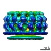
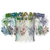
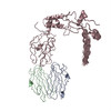
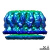
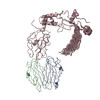
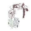
| PubMed Abstract | Membrane attack complex/perforin-like (MACPF) proteins comprise the largest superfamily of pore-forming proteins, playing crucial roles in immunity and pathogenesis. Soluble monomers assemble into ...Membrane attack complex/perforin-like (MACPF) proteins comprise the largest superfamily of pore-forming proteins, playing crucial roles in immunity and pathogenesis. Soluble monomers assemble into large transmembrane pores via conformational transitions that remain to be structurally and mechanistically characterised. Here we present an 11 Å resolution cryo-electron microscopy (cryo-EM) structure of the two-part, fungal toxin Pleurotolysin (Ply), together with crystal structures of both components (the lipid binding PlyA protein and the pore-forming MACPF component PlyB). These data reveal a 13-fold pore 80 Å in diameter and 100 Å in height, with each subunit comprised of a PlyB molecule atop a membrane bound dimer of PlyA. The resolution of the EM map, together with biophysical and computational experiments, allowed confident assignment of subdomains in a MACPF pore assembly. The major conformational changes in PlyB are a ∼70° opening of the bent and distorted central β-sheet of the MACPF domain, accompanied by extrusion and refolding of two α-helical regions into transmembrane β-hairpins (TMH1 and TMH2). We determined the structures of three different disulphide bond-trapped prepore intermediates. Analysis of these data by molecular modelling and flexible fitting allows us to generate a potential trajectory of β-sheet unbending. The results suggest that MACPF conformational change is triggered through disruption of the interface between a conserved helix-turn-helix motif and the top of TMH2. Following their release we propose that the transmembrane regions assemble into β-hairpins via top down zippering of backbone hydrogen bonds to form the membrane-inserted β-barrel. The intermediate structures of the MACPF domain during refolding into the β-barrel pore establish a structural paradigm for the transition from soluble monomer to pore, which may be conserved across the whole superfamily. The TMH2 region is critical for the release of both TMH clusters, suggesting why this region is targeted by endogenous inhibitors of MACPF function. |
 External links External links | PLoS Biol / PubMed:25654333 / PubMed Central |
| Methods | EM (single particle) / X-ray diffraction |
| Resolution | 1.85 - 17.0 Å |
| Structure data | EMDB-2793, PDB-4v2t: EMDB-2794, PDB-4v3a: EMDB-2795, PDB-4v3m: EMDB-2796, PDB-4v3n: 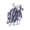 PDB-4oeb:  PDB-4oej:  PDB-4ov8: |
| Chemicals |  ChemComp-SO4:  ChemComp-HOH:  ChemComp-ACT:  ChemComp-GOL:  ChemComp-CL: |
| Source |
|
 Keywords Keywords | MEMBRANE BINDING PROTEIN / beta-sandwich fold / pleurotolysin B / MACPF domain / Pore formation / pleurotolysin A / TOXIN / TMH1-lock / pore-forming protein / Pleurtolysin A component / TRANSPORT PROTEIN / MACPF/CDC SUPERFAMILY / PORE-FORMING PROTEINS |


 pleurotus ostreatus (oyster mushroom)