 Movie
Movie Controller
 Structure viewers
 About Yorodumi Papers


+Search query
-Structure paper
| Title | ATPase cycle of the nonmotile kinesin NOD allows microtubule end tracking and drives chromosome movement. |
|---|---|
| Journal, issue, pages | Cell, Vol. 136, Issue 1, Page 110-122, Year 2009 |
| Publish date | Jan 9, 2009 |
 Authors Authors | Jared C Cochran / Charles V Sindelar / Natasha K Mulko / Kimberly A Collins / Stephanie E Kong / R Scott Hawley / F Jon Kull /  |
| PubMed Abstract | Segregation of nonexchange chromosomes during Drosophila melanogaster meiosis requires the proper function of NOD, a nonmotile kinesin-10. We have determined the X-ray crystal structure of the NOD ...Segregation of nonexchange chromosomes during Drosophila melanogaster meiosis requires the proper function of NOD, a nonmotile kinesin-10. We have determined the X-ray crystal structure of the NOD catalytic domain in the ADP- and AMPPNP-bound states. These structures reveal an alternate conformation of the microtubule binding region as well as a nucleotide-sensitive relay of hydrogen bonds at the active site. Additionally, a cryo-electron microscopy reconstruction of the nucleotide-free microtubule-NOD complex shows an atypical binding orientation. Thermodynamic studies show that NOD binds tightly to microtubules in the nucleotide-free state, yet other nucleotide states, including AMPPNP, are weakened. Our pre-steady-state kinetic analysis demonstrates that NOD interaction with microtubules occurs slowly with weak activation of ADP product release. Upon rapid substrate binding, NOD detaches from the microtubule prior to the rate-limiting step of ATP hydrolysis, which is also atypical for a kinesin. We propose a model for NOD's microtubule plus-end tracking that drives chromosome movement. |
 External links External links | Cell / PubMed:19135893 / PubMed Central |
| Methods | EM (helical sym.) / X-ray diffraction / EM (single particle) |
| Resolution | 1.9 - 11.0 Å |
| Structure data | EMDB-5038: 11-Angstrom cryo-EM reconstruction of nucleotide-free Nod complexed to the 13-protofilament microtubule. 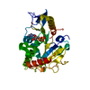 PDB-3dc4: 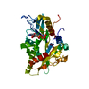 PDB-3dcb: |
| Chemicals |  ChemComp-MG:  ChemComp-ADP:  ChemComp-HOH:  ChemComp-ANP:  ChemComp-ZN:  ChemComp-GTP:  ChemComp-GDP:  ChemComp-TA1: |
| Source |
|
 Keywords Keywords |  MOTOR PROTEIN / kinesin / catalytic domain / ATPase / microtubule / ADP / nucleotide-binding protein / ATP-binding / Coiled coil / Nucleotide-binding / AMPPNP / cryo-EM / 3D reconstruction / tubulin / nucleotide-free MOTOR PROTEIN / kinesin / catalytic domain / ATPase / microtubule / ADP / nucleotide-binding protein / ATP-binding / Coiled coil / Nucleotide-binding / AMPPNP / cryo-EM / 3D reconstruction / tubulin / nucleotide-free |


