[English] 日本語
Yorodumi Papers
- Database of articles cited by EMDB/PDB/SASBDB data -

+
Search query

Keywords
Structure methods
Author
Journal
IF

-
Structure paper

TitleQueen bee pheromone binding protein pH-induced domain swapping favors pheromone release
Journal, issue, pagesJ. Mol. Biol., Vol. 390, Page 981-990, Year 2009
Publish dateApr 27, 2008 (structure data deposition date)
AuthorsPesenti, M.E. / Spinelli, S. / Bezirard, V. / Briand, L. / Pernollet, J.C. / Campanacci, V. / Tegoni, M. / Cambillau, C.
External linksJ. Mol. Biol. / PubMed:19481550
MethodsX-ray diffraction
Resolution1.5 - 2.5 Å
Structure data

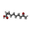
PDB-3cyz:
Dimeric crystal structure of a pheromone binding protein from Apis mellifera in complex with 9-keto-2(E)-decenoic acid at pH 7.0
Method: X-RAY DIFFRACTION / Resolution: 1.8 Å

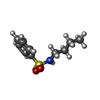
PDB-3cz0:
Dimeric crystal structure of a pheromone binding protein from Apis mellifera in complex with the n-butyl benzene sulfonamide at pH 7.0
Method: X-RAY DIFFRACTION / Resolution: 1.7 Å

PDB-3cz1:
Dimeric crystal structure of a pheromone binding protein from Apis mellifera in complex with the n-butyl benzene sulfonamide at pH 7.0
Method: X-RAY DIFFRACTION / Resolution: 1.5 Å

PDB-3cz2:
Dimeric crystal structure of a pheromone binding protein from Apis mellifera at pH 7.0
Method: X-RAY DIFFRACTION / Resolution: 2.5 Å

PDB-3d73:
Crystal structure of a pheromone binding protein mutant D35A, from Apis mellifera, at pH 7.0
Method: X-RAY DIFFRACTION / Resolution: 2.03 Å

PDB-3d74:
Crystal structure of a pheromone binding protein mutant D35A, from Apis mellifera, soaked at pH 5.5
Method: X-RAY DIFFRACTION / Resolution: 2.1 Å

PDB-3d75:
Crystal structure of a pheromone binding protein mutant D35N, from Apis mellifera, at pH 5.5
Method: X-RAY DIFFRACTION / Resolution: 2.3 Å

PDB-3d76:
Crystal structure of a pheromone binding protein mutant D35N, from Apis mellifera, soaked at pH 7.0
Method: X-RAY DIFFRACTION / Resolution: 1.9 Å

PDB-3d77:
Crystal structure of a pheromone binding protein mutant D35N, from Apis mellifera, soaked at pH 4.0
Method: X-RAY DIFFRACTION / Resolution: 1.7 Å

PDB-3d78:
Dimeric crystal structure of a pheromone binding protein mutant D35N, from apis mellifera, at pH 7.0
Method: X-RAY DIFFRACTION / Resolution: 1.6 Å

Chemicals

ChemComp-9OD:
(2Z)-9-oxodec-2-enoic acid

ChemComp-CL:
Unknown entry / Chloride

ChemComp-MG:
Unknown entry

ChemComp-GOL:
GLYCEROL / Glycerol

ChemComp-HOH:
WATER / Water

ChemComp-NBB:
N-BUTYL-BENZENESULFONAMIDE

ChemComp-NA:
Unknown entry

ChemComp-EDO:
1,2-ETHANEDIOL / Ethylene glycol

Source
  • apis mellifera (honey bee)
KeywordsPheromone Binding Protein / honeybee / Apis mellifera / signal transduction / queen mandibular pheromone / Honey bee / queen mandibular protein / pH

+
About Yorodumi Papers

-
News

-
Feb 9, 2022. New format data for meta-information of EMDB entries

New format data for meta-information of EMDB entries

  • Version 3 of the EMDB header file is now the official format.
  • The previous official version 1.9 will be removed from the archive.

Related info.:EMDB header

External links:wwPDB to switch to version 3 of the EMDB data model

-
Aug 12, 2020. Covid-19 info

Covid-19 info

URL: https://pdbj.org/emnavi/covid19.php

New page: Covid-19 featured information page in EM Navigator.

Related info.:Covid-19 info / Mar 5, 2020. Novel coronavirus structure data

+
Mar 5, 2020. Novel coronavirus structure data

Novel coronavirus structure data

Related info.:Yorodumi Speices / Aug 12, 2020. Covid-19 info

External links:COVID-19 featured content - PDBj / Molecule of the Month (242):Coronavirus Proteases

+
Jan 31, 2019. EMDB accession codes are about to change! (news from PDBe EMDB page)

EMDB accession codes are about to change! (news from PDBe EMDB page)

  • The allocation of 4 digits for EMDB accession codes will soon come to an end. Whilst these codes will remain in use, new EMDB accession codes will include an additional digit and will expand incrementally as the available range of codes is exhausted. The current 4-digit format prefixed with “EMD-” (i.e. EMD-XXXX) will advance to a 5-digit format (i.e. EMD-XXXXX), and so on. It is currently estimated that the 4-digit codes will be depleted around Spring 2019, at which point the 5-digit format will come into force.
  • The EM Navigator/Yorodumi systems omit the EMD- prefix.

Related info.:Q: What is EMD? / ID/Accession-code notation in Yorodumi/EM Navigator

External links:EMDB Accession Codes are Changing Soon! / Contact to PDBj

+
Jul 12, 2017. Major update of PDB

Major update of PDB

  • wwPDB released updated PDB data conforming to the new PDBx/mmCIF dictionary.
  • This is a major update changing the version number from 4 to 5, and with Remediation, in which all the entries are updated.
  • In this update, many items about electron microscopy experimental information are reorganized (e.g. em_software).
  • Now, EM Navigator and Yorodumi are based on the updated data.

External links:wwPDB Remediation / Enriched Model Files Conforming to OneDep Data Standards Now Available in the PDB FTP Archive

-
Yorodumi Papers

Database of articles cited by EMDB/PDB/SASBDB data

  • Database of articles cited by EMDB, PDB, and SASBDB entries
  • Using PubMed data

Related info.:EMDB / PDB / SASBDB / Yorodumi / EMN Papers / Changes in new EM Navigator and Yorodumi

Read more