 Movie
Movie Controller
 Structure viewers
 About Yorodumi Papers


+Search query
-Structure paper
| Title | A human ubiquitin conjugating enzyme (E2)-HECT E3 ligase structure-function screen. |
|---|---|
| Journal, issue, pages | Mol Cell Proteomics, Vol. 11, Page 329-341, Year 2012 |
| Publish date | Dec 6, 2004 (structure data deposition date) |
 Authors Authors | Sheng, Y. / Hong, J.H. / Doherty, R. / Srikumar, T. / Shloush, J. / Avvakumov, G.V. / Walker, J.R. / Xue, S. / Neculai, D. / Wan, J.W. ...Sheng, Y. / Hong, J.H. / Doherty, R. / Srikumar, T. / Shloush, J. / Avvakumov, G.V. / Walker, J.R. / Xue, S. / Neculai, D. / Wan, J.W. / Kim, S.K. / Arrowsmith, C.H. / Raught, B. / Dhe-Paganon, S. |
 External links External links | Mol Cell Proteomics / PubMed:22496338 |
| Methods | X-ray diffraction |
| Resolution | 1.6 - 2.56 Å |
| Structure data |  PDB-1y6l: 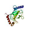 PDB-1yh2:  PDB-1yrv: 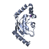 PDB-1zdn:  PDB-1zuo: 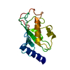 PDB-2a4d:  PDB-2a7l:  PDB-2awf:  PDB-2f4w:  PDB-2ob4:  PDB-2qgx:  PDB-2z5d:  PDB-3bzh: 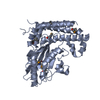 PDB-3ceg: |
| Chemicals |  ChemComp-HOH:  ChemComp-NA:  ChemComp-BME:  ChemComp-GOL: |
| Source |
|
 Keywords Keywords | LIGASE / Structural Genomics Consortium / UBIQUITIN / UBIQUITIN-CONJUGATING ENZYME / SGC / HSCP150 / STRUCTURAL GENOMICS CONSORTIUM; SGC; UBIQUITIN; UBIQUITIN-CONJUGATING ENZYME; LIGASE / Alternative splicing; Nuclear protein; Ubl conjugation pathway / UBIQUITIN- CONJUGATING ENZYME / Structural Genomics / (SGC) / Ubl conjugation pathway / Endoplasmic reticulum / Structural Genomics Consortium (SGC) / LIGASE; UBL CONJUGATION PATHWAY; STRUCTURAL GENOMICS CONSORTIUM / ubiquitin conjugating protein / UBC / UBE2H / APOPTOSIS / PROTEASE INHIBITOR / THIOL PROTEASE INHIBITOR |
 homo sapiens (human)