 Movie
Movie Controller
 Structure viewers
 About Yorodumi Papers


+Search query
-Structure paper
| Title | Crystal structures of human cardiac beta-myosin II S2-Delta provide insight into the functional role of the S2 subfragment. |
|---|---|
| Journal, issue, pages | Proc Natl Acad Sci U S A, Vol. 103, Issue 47, Page 17713-17717, Year 2006 |
| Publish date | Nov 21, 2006 |
 Authors Authors | Wulf Blankenfeldt / Nicolas H Thomä / John S Wray / Mathias Gautel / Ilme Schlichting /  |
| PubMed Abstract | Myosin II is the major component of the muscle thick filament. It consists of two N-terminal S1 subfragments ("heads") connected to a long dimeric coiled-coil rod. The rod is in itself twofold ...Myosin II is the major component of the muscle thick filament. It consists of two N-terminal S1 subfragments ("heads") connected to a long dimeric coiled-coil rod. The rod is in itself twofold symmetric, but in the filament, the two heads point away from the filament surface and are therefore not equivalent. This breaking of symmetry requires the initial section of the rod, subfragment 2 (S2), to be relatively flexible. S2 is an important functional element, involved in various mechanisms by which the activity of smooth and striated muscle is regulated. We have determined crystal structures of the 126 N-terminal residues of S2 from human cardiac beta-myosin II (S2-Delta), of both WT and the disease-associated E924K mutant. S2-Delta is a straight parallel dimeric coiled coil, but the N terminus of one chain is disordered in WT-S2-Delta due to crystal contacts, indicative of unstable local structure. Bulky noncanonical side chains pack into a/d positions of S2-Delta's N terminus, leading to defined local asymmetry and axial stagger, which could induce nonequivalence of the S1 subfragments. Additionally, S2 possesses a conserved charge distribution with three prominent rings of negative potential within S2-Delta, the first of which may provide a binding interface for the "blocked head" of smooth muscle myosin in the OFF state. The observation that many disease-associated mutations affect the second negatively charged ring further suggests that charge interactions play an important role in regulation of cardiac muscle activity through myosin-binding protein C. |
 External links External links | Proc Natl Acad Sci U S A / PubMed:17095604 / PubMed Central |
| Methods | X-ray diffraction |
| Resolution | 2.5 - 2.7 Å |
| Structure data | 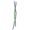 PDB-2fxm: 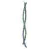 PDB-2fxo: |
| Chemicals |  ChemComp-HG: |
| Source |
|
 Keywords Keywords | CONTRACTILE PROTEIN / COILED COIL (DIMERIC / PARALLEL) / FAMILIAL HYPERTROPHIC CARDIOMYOPATHY / THICK FILAMENT / FHC-ASSOCIATED MUTANT E924K |
 homo sapiens (human)