 Movie
Movie Controller
 Structure viewers
 About Yorodumi Papers


+Search query
-Structure paper
| Title | Electron-crystallographic refinement of the structure of bacteriorhodopsin. |
|---|---|
| Journal, issue, pages | J Mol Biol, Vol. 259, Issue 3, Page 393-421, Year 1996 |
| Publish date | Jun 14, 1996 |
 Authors Authors | N Grigorieff / T A Ceska / K H Downing / J M Baldwin / R Henderson /  |
| PubMed Abstract | Using electron diffraction data corrected for diffuse scattering together with additional phase information from 30 new images of tilted specimens, an improved experimental density map has been ...Using electron diffraction data corrected for diffuse scattering together with additional phase information from 30 new images of tilted specimens, an improved experimental density map has been calculated for bacteriorhodopsin. The atomic model has then been rebuilt into this new map with particular attention to the surface loops. All the residues from 7 to 227 as well as ten lipid molecules are now included, although a few amino acid residues in three of the six surface loops, about half of the lipid hydrophobic chains and all of the lipid head groups are disordered. The model has then been refined against the experimental diffraction amplitudes to an R-factor of 28% at 3.5 angstrom resolution with strict geometry (0.005 angstrom) bond length deviation) using the improvement of the "free" phase residual between calculated and experimental phases from images as an objective criterion of accuracy. For the refinement some new programs were developed to restrain the number of parameters, to be compatible with the limited resolution of our data. In the final refined model of the protein (2BRD), compared with earlier co-ordinates (1BRD), helix D has been moved towards the cytoplasm by almost 4 angstrom, and the overall accuracy of the co-ordinates of residues in the other six helices has been improved. As a result the positions of nearly all the important residues in bacteriorhodopsin are now well determined. In particular, the buried, protonated Asp115 is 7 angstrom from, and so not in contact with, the retinal and Met118 forms a cap on the pocket occupied by the beta-ionone ring. No clear density exists for the side-chain of Arg82, which forms a central part of the extracellular half-channel. The only arginine side-chain built into good density is that of Arg134 at the extracellular end of helix E, the others being disordered near one of the two surfaces. The interpretation of the end of helix F on the extracellular surface is now clearer; an extra loose helical turn has been built bringing the side-chain of Glu194 close to Arg134 to form a probable salt bridge. The model provides an improved framework for understanding the mechanism of the light-driven proton pumping. A number of cavities that could contain water molecules were found by searching the refined model, most of them above or below the Schiff base in the half-channels leading to the two surfaces. The ordered and disordered regions of the structure are described by the temperature factor distribution. |
 External links External links | J Mol Biol / PubMed:8676377 |
| Methods | EM (electron crystallography) |
| Resolution | 3.5 Å |
| Structure data | 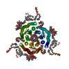 PDB-2brd: |
| Chemicals |  ChemComp-DPG:  ChemComp-RET: |
| Source |
|
 Keywords Keywords | PHOTORECEPTOR / PROTON PUMP / MEMBRANE PROTEIN / RETINAL PROTEIN / TWO-DIMENSIONAL CRYSTAL |
 halobacterium salinarum (Halophile)