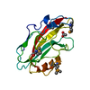
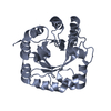
PDB-1o60 KDO-8-phosphate synthaseMethod : X-RAY DIFFRACTION / Resolution : 1.8 Å
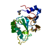
PDB-1o61 enzyme with PLP Method : X-RAY DIFFRACTION / Resolution : 1.9 Å
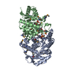
PDB-1o62 the apo form of a PLP-dependent enzymeMethod : X-RAY DIFFRACTION / Resolution : 2.1 Å
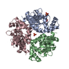
PDB-1o63 ATP phosphoribosyltransferase Method : X-RAY DIFFRACTION / Resolution : 2 Å
PDB-1o64 ATP phosphoribosyltransferase Method : X-RAY DIFFRACTION / Resolution : 2.1 Å
PDB-1o65 hypothetical proteinMethod : X-RAY DIFFRACTION / Resolution : 2.33 Å
PDB-1o66 3-methyl-2-oxobutanoate hydroxymethyltransferase Method : X-RAY DIFFRACTION / Resolution : 1.75 Å
PDB-1o67 hypothetical proteinMethod : X-RAY DIFFRACTION / Resolution : 2.54 Å
PDB-1o68 3-methyl-2-oxobutanoate hydroxymethyltransferase Method : X-RAY DIFFRACTION / Resolution : 2.1 Å
PDB-1o69 PLP-dependent enzyme Method : X-RAY DIFFRACTION / Resolution : 1.84 Å
PDB-1o6b phosphopantetheine adenylyltransferase with ADP Method : X-RAY DIFFRACTION / Resolution : 2.2 Å
PDB-1o6c UDP-N-acetylglucosamine 2-epimerase Method : X-RAY DIFFRACTION / Resolution : 2.9 Å
PDB-1o6d hypothetical proteinMethod : X-RAY DIFFRACTION / Resolution : 1.66 Å
PDB-1vgt 4-diphosphocytidyl-2C-methyl-D-erythritol synthaseMethod : X-RAY DIFFRACTION / Resolution : 1.8 Å
PDB-1vgu 4-diphosphocytidyl-2C-methyl-D-erythritol synthaseMethod : X-RAY DIFFRACTION / Resolution : 2.8 Å
PDB-1vgv UDP-N-acetylglucosamine_2 epimerase Method : X-RAY DIFFRACTION / Resolution : 2.31 Å
PDB-1vgw 4-diphosphocytidyl-2C-methyl-D-erythritol synthaseMethod : X-RAY DIFFRACTION / Resolution : 2.35 Å
PDB-1vgx autoinducer-2 synthesis proteinMethod : X-RAY DIFFRACTION / Resolution : 1.9 Å
PDB-1vgy succinyl diaminopimelate desuccinylase Method : X-RAY DIFFRACTION / Resolution : 1.9 Å
PDB-1vgz 4-diphosphocytidyl-2C-methyl-D-erythritol synthaseMethod : X-RAY DIFFRACTION / Resolution : 3 Å
PDB-1vh0 hypothetical proteinMethod : X-RAY DIFFRACTION / Resolution : 2.31 Å
PDB-1vh1 CMP-KDO synthetase Method : X-RAY DIFFRACTION / Resolution : 2.6 Å
PDB-1vh2 autoinducer-2 synthesis proteinMethod : X-RAY DIFFRACTION / Resolution : 2 Å
PDB-1vh3 CMP-KDO synthetase Method : X-RAY DIFFRACTION / Resolution : 2.7 Å
PDB-1vh4 stabilizer of iron transporter Method : X-RAY DIFFRACTION / Resolution : 1.75 Å
PDB-1vh5 thioesterase Method : X-RAY DIFFRACTION / Resolution : 1.34 Å
PDB-1vh6 flagellar proteinMethod : X-RAY DIFFRACTION / Resolution : 2.5 Å
PDB-1vh7 cyclase subunit of imidazolglycerolphosphate synthaseMethod : X-RAY DIFFRACTION / Resolution : 1.9 Å
PDB-1vh8 2C-methyl-D-erythritol 2,4-cyclodiphosphate synthaseMethod : X-RAY DIFFRACTION / Resolution : 2.35 Å
PDB-1vh9 thioesterase Method : X-RAY DIFFRACTION / Resolution : 2.15 Å
PDB-1vha 2C-methyl-D-erythritol 2,4-cyclodiphosphate synthaseMethod : X-RAY DIFFRACTION / Resolution : 2.35 Å
PDB-1vhc KHG/KDPG aldolase Method : X-RAY DIFFRACTION / Resolution : 1.89 Å
PDB-1vhd iron containing alcohol dehydrogenase Method : X-RAY DIFFRACTION / Resolution : 1.6 Å
PDB-1vhe aminopeptidase/glucanase homolog Method : X-RAY DIFFRACTION / Resolution : 1.9 Å
PDB-1vhf periplasmic divalent cation tolerance proteinMethod : X-RAY DIFFRACTION / Resolution : 1.54 Å
PDB-1vhg compounds hydrolase Method : X-RAY DIFFRACTION / Resolution : 2.7 Å
PDB-1vhj phosphorylase Method : X-RAY DIFFRACTION / Resolution : 2.23 Å
PDB-1vhk hypothetical proteinMethod : X-RAY DIFFRACTION / Resolution : 2.6 Å
PDB-1vhl adenosine-5'-diphosphate Method : X-RAY DIFFRACTION / Resolution : 1.65 Å
PDB-1vhm hypothetical proteinMethod : X-RAY DIFFRACTION / Resolution : 2.1 Å
PDB-1vho peptidase/endoglucanase Method : X-RAY DIFFRACTION / Resolution : 1.86 Å
PDB-1vhq enhancing lycopene biosynthesis protein 2 Method : X-RAY DIFFRACTION / Resolution : 1.65 Å
PDB-1vhs phosphinothricin N-acetyltransferase Method : X-RAY DIFFRACTION / Resolution : 1.8 Å
PDB-1vht bis(adenosine)-5'-triphosphate Method : X-RAY DIFFRACTION / Resolution : 1.59 Å
PDB-1vhu phosphoesterase Method : X-RAY DIFFRACTION / Resolution : 1.34 Å
PDB-1vhv diphthine synthaseMethod : X-RAY DIFFRACTION / Resolution : 1.75 Å
PDB-1vhw phosphorylase with adenosine Method : X-RAY DIFFRACTION / Resolution : 1.54 Å
PDB-1vhx Holliday junction resolvase Method : X-RAY DIFFRACTION / Resolution : 1.96 Å
PDB-1vhy Haemophilus influenzae protein HI0303, Pfam DUF558 Method : X-RAY DIFFRACTION / Resolution : 1.9 Å
PDB-1vhz compounds hydrolase Method : X-RAY DIFFRACTION / Resolution : 2.32 Å
PDB-1vi0 transcriptional regulator Method : X-RAY DIFFRACTION / Resolution : 1.65 Å
PDB-1vi1 fatty acid/phospholipid synthesis proteinMethod : X-RAY DIFFRACTION / Resolution : 2.95 Å
PDB-1vi2 shikimate-5-dehydrogenase with NAD Method : X-RAY DIFFRACTION / Resolution : 2.1 Å
PDB-1vi3 hypothetical proteinMethod : X-RAY DIFFRACTION / Resolution : 1.76 Å
PDB-1vi4 Regulator of ribonuclease activity A protein 1 Method : X-RAY DIFFRACTION / Resolution : 1.87 Å
PDB-1vi5 ribosomal protein S2P Method : X-RAY DIFFRACTION / Resolution : 2.65 Å
PDB-1vi6 ribosomal protein S2P Method : X-RAY DIFFRACTION / Resolution : 1.95 Å
PDB-1vi8 thioesterase Method : X-RAY DIFFRACTION / Resolution : 2.2 Å
PDB-1vi9 pyridoxamine kinaseMethod : X-RAY DIFFRACTION / Resolution : 1.96 Å
PDB-1via shikimate kinaseMethod : X-RAY DIFFRACTION / Resolution : 1.57 Å
PDB-1vic CMP-KDO synthetase Method : X-RAY DIFFRACTION / Resolution : 1.8 Å
PDB-1vim hypothetical proteinMethod : X-RAY DIFFRACTION / Resolution : 1.36 Å
PDB-1viq ADP ribose pyrophosphatase Method : X-RAY DIFFRACTION / Resolution : 2.4 Å
PDB-1vis mevalonate kinaseMethod : X-RAY DIFFRACTION / Resolution : 2.69 Å
PDB-1viu ADP ribose pyrophosphatase Method : X-RAY DIFFRACTION / Resolution : 2.4 Å
PDB-1viv hypothetical proteinMethod : X-RAY DIFFRACTION / Resolution : 2.6 Å
PDB-1vix peptidase T Method : X-RAY DIFFRACTION / Resolution : 2.5 Å
PDB-1viy dephospho-CoA kinaseMethod : X-RAY DIFFRACTION / Resolution : 1.89 Å
PDB-1viz hypothetical proteinMethod : X-RAY DIFFRACTION / Resolution : 1.85 Å
 Movie
Movie Controller
 Structure viewers
 About Yorodumi Papers


 Authors
 External links


 Keywords
 haemophilus influenzae (bacteria)
 campylobacter jejuni (Campylobacter)
 thermotoga maritima (bacteria)
 staphylococcus aureus (bacteria)
 vibrio cholerae (bacteria)
