 Movie
Movie Controller
 Structure viewers
 About Yorodumi Papers


+Search query
-Structure paper
| Title | Visualization of a water-selective pore by electron crystallography in vitreous ice. |
|---|---|
| Journal, issue, pages | Proc Natl Acad Sci U S A, Vol. 98, Issue 4, Page 1398-1403, Year 2001 |
| Publish date | Feb 13, 2001 |
 Authors Authors | G Ren / V S Reddy / A Cheng / P Melnyk / A K Mitra /  |
| PubMed Abstract | The water-selective pathway through the aquaporin-1 membrane channel has been visualized by fitting an atomic model to a 3.7-A resolution three-dimensional density map. This map was determined by ...The water-selective pathway through the aquaporin-1 membrane channel has been visualized by fitting an atomic model to a 3.7-A resolution three-dimensional density map. This map was determined by analyzing images and electron diffraction patterns of lipid-reconstituted two-dimensional crystals of aquaporin-1 preserved in vitrified buffer in the absence of any additive. The aqueous pathway is characterized by a size-selective pore that is approximately 4.0 +/- 0.5A in diameter, spans a length of approximately 18A, and bends by approximately 25 degrees as it traverses the bilayer. This narrow pore is connected by wide, funnel-shaped openings at the extracellular and cytoplasmic faces. The size-selective pore is outlined mostly by hydrophobic residues, resulting in a relatively inert pathway conducive to diffusion-limited water flow. The apex of the curved pore is close to the locations of the in-plane pseudo-2-fold symmetry axis that relates the N- and C-terminal halves and the conserved, functionally important N76 and N192 residues. |
 External links External links | Proc Natl Acad Sci U S A / PubMed:11171962 / PubMed Central |
| Methods | EM (electron crystallography) |
| Resolution | 3.7 Å |
| Structure data | 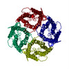 PDB-1ih5: |
| Source |
|
 Keywords Keywords | MEMBRANE PROTEIN / WATER CHANNEL / TWO-DIMENSIONAL CRYSTAL |
 homo sapiens (human)