 Movie
Movie Controller
 Structure viewers
 About Yorodumi Papers


+Search query
-Structure paper
| Title | Structural studies of two rhinovirus serotypes complexed with fragments of their cellular receptor. |
|---|---|
| Journal, issue, pages | EMBO J, Vol. 18, Issue 22, Page 6249-6259, Year 1999 |
| Publish date | Nov 15, 1999 |
 Authors Authors | P R Kolatkar / J Bella / N H Olson / C M Bator / T S Baker / M G Rossmann /  |
| PubMed Abstract | Two human rhinovirus serotypes complexed with two- and five-domain soluble fragments of the cellular receptor, intercellular adhesion molecule-1, have been investigated by X-ray crystallographic ...Two human rhinovirus serotypes complexed with two- and five-domain soluble fragments of the cellular receptor, intercellular adhesion molecule-1, have been investigated by X-ray crystallographic analyses of the individual components and by cryo-electron microscopy of the complexes. The three-dimensional image reconstructions provide a molecular envelope within which the crystal structures of the viruses and the receptor fragments can be positioned with accuracy. The N-terminal domain of the receptor binds to the rhinovirus 'canyon' surrounding the icosahedral 5-fold axes. Fitting of molecular models into the image reconstruction density identified the residues on the virus that interact with those on the receptor surface, demonstrating complementarity of the electrostatic patterns for the tip of the N-terminal receptor domain and the floor of the canyon. The complexes seen in the image reconstructions probably represent the first stage of a multistep binding process. A mechanism is proposed for the subsequent viral uncoating process. |
 External links External links | EMBO J / PubMed:10562537 / PubMed Central |
| Methods | EM (single particle) / X-ray diffraction |
| Resolution | 3.25 - 28 Å |
| Structure data |  PDB-1d3e: 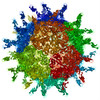 PDB-1d3i:  PDB-1d3l: |
| Source |
|
 Keywords Keywords | Virus/Receptor /  HUMAN RHINOVIRUS / HRV16 / ICAM-1 / FITTING OF X-RAY STRUCTURES INTO CRYO-EM RECONSTRUCTIONS / COMMON COLD / VIRUS UNCOATING / VIRUS/ VIRAL PROTEIN / RHINOVIRUS-RECEPTOR COMPLEX / Icosahedral virus / Virus-Receptor COMPLEX / HRV14 / CELL ADHESION / RHINOVIRUS RECEPTOR / ADHESION PROTEIN / GLYCOPROTEIN / IMMUNOGLOBULIN FOLD HUMAN RHINOVIRUS / HRV16 / ICAM-1 / FITTING OF X-RAY STRUCTURES INTO CRYO-EM RECONSTRUCTIONS / COMMON COLD / VIRUS UNCOATING / VIRUS/ VIRAL PROTEIN / RHINOVIRUS-RECEPTOR COMPLEX / Icosahedral virus / Virus-Receptor COMPLEX / HRV14 / CELL ADHESION / RHINOVIRUS RECEPTOR / ADHESION PROTEIN / GLYCOPROTEIN / IMMUNOGLOBULIN FOLD |


